Fig 3 and 4 give the obtained patient effective dose levels in case of the regular scan mode, showing the results for the dento-maxillofacial/ENT and extremity imaging applications, respectively. Fig 5 and 6 display the results for the best scan mode.
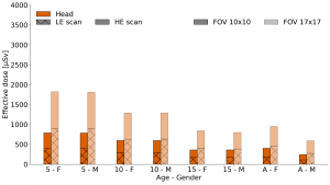
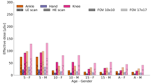
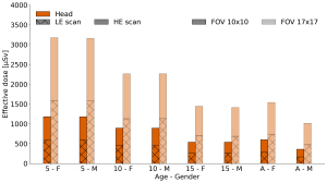
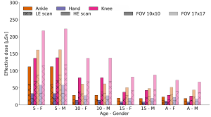
For all the studied protocols, a decreasing trend as a function of patient age could be observed. Most often, similar dose levels were obtained for the 15 year old and adult patient models. In addition, both the LE and HE scan contributed about equally to the total dose. In general, lower dose levels were observed for the extremity imaging protocols than for the dento-maxillofacial/ENT imaging applications. This can be explained by the presence of more radiosensitive organs in the head region. For the extremity imaging applications, a knee scan resulted in the highest dose level, followed by ankle and hand. As expected from the tube-current exposure time values given in Table 1, patient dose levels increased for the best scan mode compared to the regular one. Furthermore, dose levels increased with increasing FOV size, for the same scan mode.
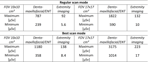
Table 2 provides an overview of the maximum and minimum dose values (µSv) for the studied imaging protocols, for both the dento-maxillofacial/ENT and extremity imaging applications.
Overall, dose levels can be considered acceptable for the applications studied, especially since this is, to the author’s best knowledge, the first commercially available model on the European market. Future studies, comparing the obtained values to conventional CBCT and CT as well as DE CT imaging for similar clinical applications will allow to situate the system better in terms of patient dose levels. In addition, official introduction of the system into general clinical practice, will possibly enable to further investigate patient dose levels, and set-up clinical application specific dose optimization studies.