Back to the list
Congress: ECR25
Poster Number: C-23680
Type: Poster: EPOS Radiologist (scientific)
Authorblock: S. M. R. S. Islam1, A. Biguri2, C. Landi3, G. Di Domenico4, P. Grün5, D. Turhani5, G. Kronreif1, W. Birkfellner6, S. Hatamikia7; 1Wiener Neustadt/AT, 2Cambridge/UK, 3Via Bolgara 2, Brusaporto (BG)/IT, 4Ferrara/IT, 5Steiner Landstraße 124, Krems/AT, 6Vienna/AT, 7Krems/AT
Disclosures:
S M Ragib Shahriar Islam: Nothing to disclose
Ander Biguri: Nothing to disclose
Claudio Landi: Nothing to disclose
Giovanni Di Domenico: Nothing to disclose
Pascal Grün: Nothing to disclose
Dritan Turhani: Nothing to disclose
Gernot Kronreif: Nothing to disclose
Wolfgang Birkfellner: Nothing to disclose
Sepideh Hatamikia: Nothing to disclose
Keywords: Interventional non-vascular, Cone beam CT, Digital radiography, CAD, Cost-effectiveness, Experimental investigations, Image verification, Kv imaging
Results

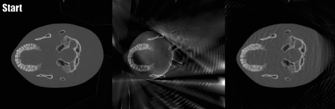
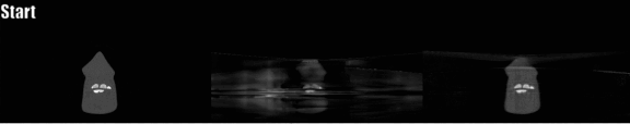
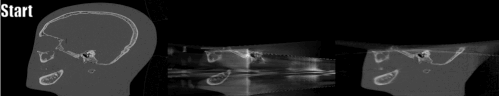
Reconstructed Images and Extended Field of View (FOV):

  • The FOV for the limited-angle circular scan (in the axial plane) was a circle with a diameter of 11 cm.
  • Using the proposed trajectory, the reconstructed volume expanded to approximately 15 cm in cranial breadth and 19.5 cm in the naso-occipital length.
  • This represents an FOV increase of approximately 77%.

Fig 7: The comparison of reconstructed images from the limited angle circular scan and the proposed fusion trajectory with the digital phantom image

The following animated graphics provide a comparative illustration of the reconstructed images obtained using limited-angle circular scan data and the proposed fusion trajectory scan data, alongside the phantom image, across different imaging planes (Axial, Coronal, and Sagittal).

Fig 8: The comparison in axial plane. (Left: The phantom image; Middle: Limited angle circular scan reconstructed image); Right: The proposed fusion trajectory scan reconstructed image

Fig 9: The comparison in coronal plane. (Left: The phantom image; Middle: Limited angle circular scan reconstructed image); Right: The proposed fusion trajectory scan reconstructed image

Fig 10: The comparison in sagittal plane. (Left: The phantom image; Middle: Limited angle circular scan reconstructed image); Right: The proposed fusion trajectory scan reconstructed image

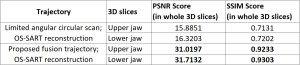
Quantitative Analysis of Extended FOV Images: To evaluate the reconstructed images from both the limited-angle circular scan and the proposed fusion trajectory, Peak Signal-to-Noise Ratio (PSNR) and Structural Similarity Index Measurement (SSIM) scores were calculated by comparing them to the digital phantom image.

  • PSNR: This metric indicates the accuracy of image reconstruction. A PSNR value of 30 dB or higher is considered indicative of good reconstruction relative to the ground truth.
  • SSIM: This metric assesses the visual quality of the image. Values closer to 1.00 represent better visual similarity to the ground truth.
  • Results: Images reconstructed using the proposed fusion trajectory achieved higher PSNR and SSIM scores, demonstrating superior reconstruction quality and visual fidelity compared to those from the limited-angle circular scan.

The following table presents the PSNR and SSIM metric scores for the reconstructed images of the upper and lower jaw volumes, comparing the limited-angle circular scan and the proposed fusion trajectory scan.

Table 11: The image quality measurement metric scores for the limited angle circular scan and the proposed fusion trajectory scan reconstructed images.


GALLERY