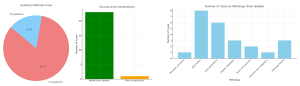
Over the past 12 months, cryoablation was performed on 24 patients with a variety of malignant and non-malignant pathologies. Of these, 1 patient was treated for a recidivant pancreatic carcinoma, 6 for lung malignancies, 3 for hepatic metastases, 8 for bone lesions, 1 for head and neck malignancy, 2 for renal cell carcinoma (stage T1A and T1B), and 3 for soft tissue lesions. The procedures were conducted under general anesthesia, with CT guidance used in 20 cases and ultrasound (US) guidance in 4 cases, with a focus on visualizing the "iceball" created during the ablation process. Post-procedural imaging and follow-up were conducted to assess treatment efficacy and monitor complications. The technical success rate of the procedures was 100%, with whole tumor ablation achieved in 23 of 24 patients. There was 1 minor complication (4.2%), and no major complications were reported.
Detailed Findings:
-
Pancreatic carcinoma: A 63-year-old woman with recurrent pancreatic malignoma three years after open surgical irreversible electroporation of the pancreatic head. The procedure was technically successful; however, a post-ablational biloma was detected, representing the only minor complication in this series.
 Fig 4: A patient with a recurrent pancreatic tumor underwent placement of a single cryoprobe, followed by iceball necrosis after two freeze-thaw cycles.
 -
Bone lesions (8 cases):
- 4 cases of osteoid osteomas (all in the femur).
- 1 osteoid osteoma located in the C3 transverse process.
- 2 metastatic rib lesions.
- 1 osteoblastoma of the L3 vertebral body.
 Fig 5: CT showing proper needle placement in a biopsy proven rib metastasis from transitional cell bladder carcinoma. FUCT after two freeze–thaw cycles showing hypodense ice ball surrounding the lesion.
 Fig 6: The first four images show MRI scans of a 19-year-old male patient with severe pain due to an osteoid osteoma in the left C3 transverse process. The initial MRI images demonstrate the lesion's location and its characteristics. The following two images depict the cryoablation procedure, with two probes placed in the core of the osteoid osteoma. The CT images show a tiny hypodense area around the soft tissue, indicating the targeted treatment area and the initial result of the cryoablation.
- Hepatic metastases:
-
- One patient had a metastasis originating from lung cancer.
- Two cases of hepatic metastases were secondary to colon carcinoma.
 Fig 7: A PET-CT scan in a patient with colon carcinoma showed positive FDG-uptake in a 15x12 mm focal hepatic lesion. Two cryoprobes, placed 7 mm apart, delivered two freeze-thaw cycles to treat the metastasis. A CT scan during the procedure revealed the iceball, measuring 35 mm, clearly visible around the target lesion.
-
Lung malignancies (6 cases):
- 3 cases of primary lung tumors.
- 3 cases of metastatic lung lesions (colon and breast carcinomas).
 Fig 8: Pre-interventional CT shows a small nodular change in the left lower lobe, which demonstrated positive FDG-uptake in a patient with colon cancer. During the procedure, the CT image reveals a hypodense area around the cryoprobe, with minimal damage to the surrounding tissue.
- Renal cell carcinoma (2 cases):
- 1 patient with stage T1A RCC.
- 1 patient with stage T1B RCC.
 Fig 9: T1fsC+ MR sequence showing a 12 mm biopsy-proven RCC in the upper pole of the right kidney. A hypodense "iceball" with a cryoablation needle, visible on computed tomography during percutaneous cryoablation of the RCC lesion (coronal reconstruction). 1-month follow-up CT showing complete necrosis and tumor ablation in the venous phase.
-
Head and neck malignancy (1 case): A single patient with a metastasis from a squamous cell carcinoma of the larynx.
-
Soft tissue lesions (3 cases):
-
- Two female patients with abdominal wall endometriosis following Caesarean section. Both patients presented with abdominal pain and menstrual cycle–related cramps.
- One patient had a metastasis from colon carcinoma involving the left psoas muscle.
 Fig 10: Percutaneous US guided cryoablation needle placement in an endometrioma in the abdominal wall. Ultrasound showing cone shadowing as an indirect visualization of the anechoic presentation of the iceball.
 Fig 11: A pre-interventional PET CT scan in a patient with colon carcinoma showed positive FDG uptake in a lesion in the left psoas muscle. A hypodense "iceball" was visible on computed tomography during percutaneous cryoablation of the same lesion, with two cryoablation needles inserted.
In this case series, the iceball was successfully visualized in all procedures, enabling precise targeting and ablation of the lesions. The procedure was well-tolerated by patients, with a complication rate of 4.2% (1 minor complication out of 24 procedures) and no major complications. Follow-up imaging confirmed effective tumor ablation in 92% of cases, demonstrating the efficacy of this technique. Additionally, there was significant improvement in symptom management, particularly in cases involving bone and soft tissue lesions. The versatility of cryoablation was evident in its application across diverse pathologies, ranging from malignant tumors to benign conditions, providing therapeutic benefit and enhancing the quality of life for patients.