
Congress:
ECR26
Poster Number:
C-10853
Type:
Poster: EPOS Radiologist (educational)
Authorblock:
J. H. B. P. Furlan, E. K. U. N. Fonseca, M. V. Y. Sawamura, P. Gaspar Dos Santos, J. M. Cortez Filho, M. Tonholo Ikedo, P. Esrom, F. D. C. Bernardi, A. S. Silva Mesquita; São Paulo/BR
Disclosures:
João Henrique Barros Penteado Furlan:
Nothing to disclose
Eduardo Kaiser Ururahy Nunes Fonseca:
Nothing to disclose
Marcio Valente Yamada Sawamura:
Nothing to disclose
Pedro Gaspar Dos Santos:
Nothing to disclose
João Martins Cortez Filho:
Nothing to disclose
Matheus Tonholo Ikedo:
Nothing to disclose
Paulo Esrom:
Nothing to disclose
Fabiola Del Carlo Bernardi:
Nothing to disclose
Ana Sofia Silva Mesquita:
Nothing to disclose
Keywords:
Mediastinum, CT, Digital radiography, MR, Diagnostic procedure, Cancer, Cysts, Pathology
To address localization inconsistencies, the International Thymic Malignancy Interest Group (ITMIG) established a standard CT-based classification (2017), dividing the mediastinum into three distinct compartments [1]. This anatomical division provides a systematic framework for differential diagnosis based on the specific contents of each zone:
- Prevascular Compartment:
- Definition: From the posterior border of the sternum to the anterior aspect of the pericardium.
- Contents: Thymus, fat, lymph nodes, and the left brachiocephalic vein.
- Visceral Compartment:
- Definition: Extending from the posterior boundary of the prevascular zone to a vertical line 1 cm posterior to the anterior margin of the thoracic vertebral bodies.
- Contents: Vascular structures (heart, aorta, SVC, pulmonary arteries), thoracic duct, and non-vascular organs (trachea, esophagus, lymph nodes).
- Paravertebral Compartment:
- Definition: The posterior-most region, extending laterally to the transverse processes.
- Contents: Thoracic spine, paravertebral soft tissues, and neural elements.
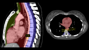
Fig 1: CT Visualization of Mediastinal Compartments (Based on ITMIG 2017 classification [1]). Sagittal overlay (Left) delineates the three distinct zones: Prevascular (yellow), Visceral (red), and Paravertebral (blue). Axial view (Right) demonstrates the Visceral (red) and Paravertebral (blue) compartments. Note that the prevascular compartment is not visible at this specific anatomical level.