Back to the list
Congress: ECR25
Poster Number: C-20900
Type: Poster: EPOS Radiologist (scientific)
Authorblock: T. Yeshua, T. Amiel, E. Halle, C. Nadler; Jerusalem/IL
Disclosures:
Talia Yeshua: Nothing to disclose
Tevel Amiel: Nothing to disclose
Elia Halle: Nothing to disclose
Chen Nadler: Nothing to disclose
Keywords: Artificial Intelligence, Head and neck, Salivary glands, Cone beam CT, CAD, Pathology
Results

The AI application demonstrated progressive improvement through the development stages. Initial validation on original MIP images (without ROI definition) achieved an accuracy of 0.91, sensitivity of 0.94, and specificity of 0.88. After implementing ROI definition, the validation performance improved significantly, reaching an accuracy of 0.97, sensitivity of 0.99, and specificity of 0.94. The final evaluation on the independent test set of 40 cases showed exceptional performance, with an accuracy of 0.99, sensitivity of 1.0, and specificity of 0.99, with a rapid processing time of 40 seconds per scan.

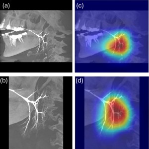
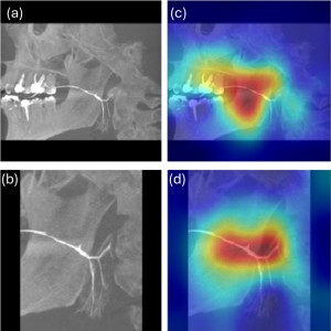
To understand the algorithm's decision-making process, GRAD-CAM analysis was performed to visualize the significant pixels contributing to classification decisions. Figure 1 demonstrates a normal case before (a) and after (b) ROI definition, with corresponding GRAD-CAM maps (c,d). Figure 2 shows a ductopenic case with similar visualization arrangement. In the normal case (Figure 1), the algorithm successfully focused on salivary ducts both in original and ROI-defined images. However, in the ductopenic case (Figure 2), the original MIP image analysis showed attention to irrelevant areas including dental restorations such as crowns and roots, while the ROI-defined analysis demonstrated more precise focus on the main salivary duct and its central branching patterns. These findings highlight the importance of proper ROI definition in achieving accurate classification.


Fig 1: GRAD-CAM visualization of a normal-appearing parotid gland. (a) Original sagittal MIP image showing complete ductal arborization; (b) MIP image after ROI definition highlighting the parotid region; (c) GRAD-CAM heat map overlay on the original MIP image demonstrating algorithm attention to ductal structures; (d) GRAD-CAM heat map overlay on the ROI-defined image showing maintained focus on salivary ducts and their branching patterns. Note the algorithm's consistent attention to the quaternary and quinary ductal branches in both original and ROI-defined images, indicating proper feature recognition regardless of image preprocessing.
Fig 2: GRAD-CAM visualization of a ductopenic parotid gland. (a) Original sagittal MIP image showing reduced ductal arborization; (b) MIP image after ROI definition focusing on the parotid region; (c) GRAD-CAM heat map overlay on the original MIP image showing algorithm attention dispersed across dental structures and non-ductal regions; (d) GRAD-CAM heat map overlay on the ROI-defined image demonstrating improved focus on the main salivary duct and its limited branching pattern. Note the significant improvement in algorithm attention after ROI definition, with focus shifted from dental artifacts to relevant ductal structures, highlighting the importance of proper image preprocessing for accurate ductopenia detection.


GALLERY