Congress:
ECR25
Poster Number:
C-23126
Type:
Poster: EPOS Radiologist (scientific)
DOI:
10.26044/ecr2025/C-23126
Authorblock:
E. Pershina, K. Kovalev, Z. Magomedova, D. Shchekochikhin; Moscow/RU
Disclosures:
Ekaterina Pershina:
Nothing to disclose
Konstantin Kovalev:
Nothing to disclose
Zaynab Magomedova:
Nothing to disclose
Dmitry Shchekochikhin:
Nothing to disclose
Keywords:
Cardiac, MR, Contrast agent-intravenous, Hyperplasia / Hypertrophy, Tissue characterisation
In this study, healthy controls, with an average age of 47.5±15.2 years, were younger than both the HCM patients, who averaged 53.9±10.5 years, and the LVH patients, who averaged 58.5±14.5 years (p=0.01). There were no significant differences in sex distribution between the groups. Several strain parameters exhibited significant differences between the HCM and LVH groups, pointing to distinct myocardial deformation patterns
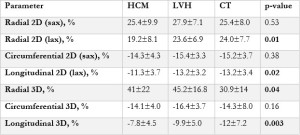
Table 1: Strain parameter differences between HCM, LVH, and healthy control groups