Using a 1.5T and 3T whole body MR scanner (Magnetom AvantoFit and Magnetom Skyra, Siemens, Erlangen, Germany) with an 18-channel breast coil artifact sizes of the four breast biopsy markers were measured in an agarose gel phantom using T2-weighted fast spin-echo imaging with short TI inversion recovery fat suppression and magnitude reconstruction (T2-TIRM), T1-weighted spoiled gradient echo sequence with fat suppression (T1_FL3D) routinely used for dynamic contrast-enhanced imaging and diffusion weighted imaging (DWI) including a readout segmented echo-planar imaging (RESOLVE-DWI) and echo planar imaging sequence (EPI-DWI).
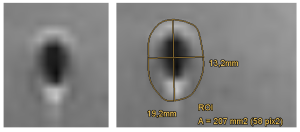
All images were analyzed using our institution’s picture archiving and communication system (PACS) software Impax EE (R20 XVII SU1, Agfa, Mortsel, Belgium). The dimensions of the visible artifact were quantified on the image with the largest appearance of the artifact by tracing the outlines of the visible signal changes on this image. The area of the obtained spline-interpolated polygonal region of interest (ROI) and the extent of the long and short axis of this ROI were recorded. Fig. 2 provides an exemplary measurement.