Back to the list
Congress: ECR24
Poster Number: C-14778
Type: EPOS Radiologist (educational)
DOI: 10.26044/ecr2024/C-14778
Authorblock: A. Calero Ortega, M. M. Merideño García, J. López Martín, E. Ponte, J. Tejedor Toquero, M. S. Paez Alvarez, E. F. Berríos, V. Esteban Izquierdo, A. D. Onuta; Toledo/ES
Disclosures:
Andrea Calero Ortega: Nothing to disclose
María Montaña Merideño García: Nothing to disclose
Jaime López Martín: Nothing to disclose
Elisabetta Ponte: Nothing to disclose
Javier Tejedor Toquero: Nothing to disclose
Manuel Sebastian Paez Alvarez: Nothing to disclose
Esnelly Francismaría Berríos: Nothing to disclose
Victoria Esteban Izquierdo: Nothing to disclose
Andrei Daniel Onuta: Nothing to disclose
Keywords: Anatomy, Respiratory system, Conventional radiography, CT, Computer Applications-3D, Computer Applications-General, Cavitation, Cysts
Background

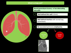
As defined by the Fleishchner Society glossary of terms, lung cysts are round parenchymal lucencies or low-attenuation areas with well-defined interfaces with normal adjacent lungs. They are characteristically thin-walled structures with walls less than 3 mm in size, usually containing air, but also fluid or solid material.

Fig 1: A true lung cyst

Isolated pulmonary parenchymal cysts are seen commonly on computed tomography, but we should be aware that there is also a wide heterogeneous group of diseases associated with lung cysts, making the differential diagnosis challenging. Thus, for radiologic assessment of cystic lung diseases, it is very important to differentiate true lung cysts from other air-space lesions (cyst-like lesions), in order to achieve an accurate diagnosis.

GALLERY