
Study Design and Population
This retrospective study included 170 patients who underwent preoperative contrast-enhanced CT between April 2017 and October 2021. Patients with bowel obstruction, inflammatory disease, or collateral venous pathways were excluded. The final cohort was divided into two groups:
- CO₂ insufflation group: n = 86.
- Non-insufflation group: n = 84.
 Fig 4: A total of 170 patients who underwent preoperative contrast-enhanced CT colonography were retrospectively analyzed and divided into CO₂ insufflation and non-insufflation groups.
CT Acquisition Protocol
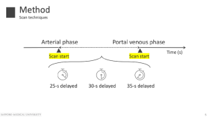
Portal venous phase imaging was performed at fixed delays of 25, 30, or 35 seconds after initiation of arterial-phase scanning. Attenuation values (HU) of the IMV and SMV were measured at their venous roots using standardized region-of-interest placement.
In the CO₂ insufflation group, colonic gas volume was quantified to reflect the degree of intraluminal CO₂ insufflation.
Statistical Analysis
The following analyses were performed:
- Comparison of venous attenuation across scan delays.
- Correlation analysis between enhancement and scan delay.
- Multivariate linear regression analysis restricted to the CO₂ insufflation group to identify predictors of mesenteric venous enhancement.
 Fig 6: CT attenuation values of the IMV and SMV were measured at the venous roots, followed by correlation analysis with scan delay and multivariate regression to identify predictors of venous enhancement.