
Congress:
ECR26
Poster Number:
C-12957
Type:
Poster: EPOS Radiologist (educational)
Authorblock:
F. Stoica1, S. L. Ghiea2; 1Timisoara/RO, 2Bucharest/RO
Disclosures:
Felicia Stoica:
Nothing to disclose
Sorin Lucian Ghiea:
Nothing to disclose
Keywords:
Bones, Musculoskeletal system, CT, MR, Structured reporting, Technical aspects, Cancer, Haematologic diseases
Bone tumours represent a heterogeneous group of diseases from both an epidemiological and histopathological perspective, and they can be broadly classified as:
(1) primary or secondary: primary bone tumours are overall rare, with secondary metastatic lesions being far more prevalent;
(2) benign, intermediate or malignant: depending on the histopathological features and biological behaviour of the tumour, with the WHO classification of bone tumours serving as a reference guide.
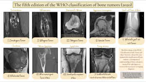
Fig 1: The fifth edition of the WHO Classification of Soft Tissue and Bone Tumours (2020), including one example of a tumour for each histopathological class.
Regarding imaging techniques, despite technological advances, radiography remains the preferred initial investigation for suspected bone tumours, highlighting benign or malignant features.
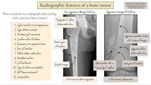
Fig 2: Radiographic features of bone tumours.