Back to the list
Congress: ECR25
Poster Number: C-23499
Type: Poster: EPOS Radiologist (scientific)
DOI: 10.26044/ecr2025/C-23499
Authorblock: A. Ben Lakhal, S. Boukriba, A. Eya, W. Frikha, H. Mizouni; Tunis/TN
Disclosures:
Amine Ben Lakhal: Nothing to disclose
Seifeddine Boukriba: Nothing to disclose
Azouz Eya: Nothing to disclose
Wassim Frikha: Nothing to disclose
Habiba Mizouni: Nothing to disclose
Keywords: Ear / Nose / Throat, Head and neck, CT, MR, Imaging sequences, Pathology
Results

All in all, we found 160 MRIs that included the late-enhanced 3D-FLAIR sequence in their protocol. Of these patients, 4 were found to have secondary endolymphatic hydrops.

In all cases, patients prestented with a triade of vertigo, tinnitus and hearing loss and the diagnosis of MD was suspected.

Case #1 was a patient who was suspected of having left-sided MD. On imaging, we found a dilated cochlear canal on the left side as well as an intravestibular schwannoma. We conluded to a secondary cochlear EH.
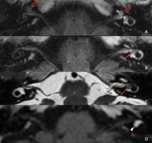
Fig 1: In A, we can see a dilated cochlear canal on the left, contrasting with a normal cochlear canal on the right (shallow arrows). In B, the intra-vestibular schwannoma can be seen as a filling defect in the vestibule. On conventional imaging, the schwannoma had an intermediate single on T2SE (C) and was homogeneously enhanced (D). An asymmetrical perilymphatic enhancement can also be noted in A and B, with a much more vivid enhancement on the left-side. This is due to the presence of the intravestibular tumor which alters the blood-perilymph barrier.

The second patient was also a suspected case of left-sided MD. On imaging, we found saccular hydrops as well as an arachnoid cyst of the cerebello-pontine angle (CPA). CPA tumors inluding arachnoid cysts have been described in the litterature in association with symptoms mimicking MD. Most believe that the pathophysiology is secondary endolymphatic hydrops, our imaging is in line with this theory.

Fig 2: In A and B, we can see the arachnoid cyst (arrows) as well-defined, thin-walled, cystic mass in the left CPA. In C, the utricle is not dilated and does not protrude in the semi-circular canal. In D, we can clearly see the saccular hydrops on the left side (arrowhead).

The third patient had previously undergone surgery for otosclerosis 4 years prior. After initial improvement, they presented with vertigo, tinnitus and mixed hearing loss. Secondary displacement of the piston-wire prothesis was suspect and a temporal bone CT was performed which came back negative. They were then further investigated using MRI which confirmed the presence of both a cochlear and a vestibular endolymphatic hydrops on the left side.
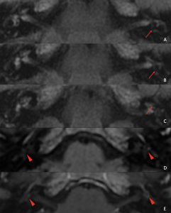
Fig 3: Figures A, B and C, show respectively (red arrows), a utricular, saccular and cochlear endolymphatic hydrops on the left side. On D, otosclerotic foci are seen as region with tissular signal (moderately low on T1). On E, these foci moderately enhance.

Patient number 4 presented with bilateral tinnitus, vertigo and hearing loss on the right side. Imaging found bilateral biletaral endolymphatic hydrops. Sequences exploring the infra and subtentorial regions showed signs of intracranial hypotension.
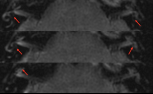
Fig 4: Bilateral endolymphatic hydrops with protrusion of the utricle in the semi-circular canal on both sides. A dilated saccule on both sides and cochlear hydrops on the right side.

Fig 5: Hallmarks of intracranial hypotension found in patient 4 with a "sagging brain" on sagittal T2 SE images (A), a reduced ponto-mesencephalic angle (B) and diffuse pachymeningeal enhancement (C).

GALLERY