
Congress:
ECR26
Poster Number:
C-11391
Type:
Poster: EPOS Radiologist (educational)
Authorblock:
I. Togrul, A. G. Erdemir, S. Arslan Sarıkaya, A. E. Yıldız; Ankara/TR
Disclosures:
Irem Togrul:
Nothing to disclose
Ahmet Gürkan Erdemir:
Nothing to disclose
Sevtap Arslan Sarıkaya:
Nothing to disclose
Adalet Elçin Yıldız:
Nothing to disclose
Keywords:
Bones, Musculoskeletal bone, Conventional radiography, CT, MR, Education, Demineralisation-Bone, Trauma
The patella is the largest sesamoid bone and lacks a true periosteum. It's located anterior to the knee, embedded within the patellar tendon, serving as an attachment site for quadriceps and patellar tendons[1,2].
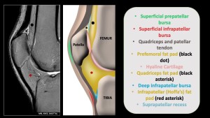
Fig 1: Patella and related anatomy
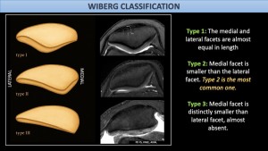
Fig 2: Wiberg classification