Imaging system and software for noise reduction
The CXDI-720C (Canon Medical Systems) was used for image acquisition. The pixel size of the images was 0.125 × 0.125 mm, with a 2800 × 3408 matrix and 16-bit grayscale. X-ray exposure was performed using an X-ray system with a total filtration of 2.8 mm Al.
For noise reduction processing, two methods available within this imaging system were used. One is a conventional rule-based approach (conventional noise reduction: Con-NR), and the other was a deep-learning-based method (Intelligent noise reduction: INR) [11,12]. These processes can be arbitrarily set to 10 levels of noise reduction intensity. For this evaluation, Levels 5 and 10 were used for both methods. The proposed method was applied to these processes to investigate their operational characteristics.
Noise characteristics evaluation of uniform images
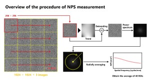
To evaluate the effect of noise reduction on uniformly exposed images, the NPS was measured using the two-dimensional Fourier transform method in accordance with the IEC 62220-1-1 [15].
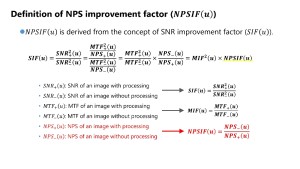
The NPSIF is defined as the ratio of the NPS before and after image processing and can be used as a factor to explain the impact on signal-to-noise ratio (SNR) improvement [16]. In this verification, for each dose condition, the NPS of images without noise reduction processing was defined as NPS-(u), and the NPS of processed images was defined as NPS+(u) for the calculation.
In this evaluation, the beam condition of RQA5 specified in IEC 61267 was employed [17]. The X-ray tube was operated at a tube-voltage of 71 kV, with a 21-mm-thick aluminum filter attached to the tube window to adjust the beam quality. The imaging distance was set to 150 cm, and imaging doses were adjusted by varying the tube-current-time product at 0.5, 1.0, and 5.0 mAs.
Noise characteristics evaluation of phantom images
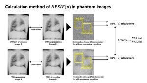
To evaluate the noise reduction effects on images containing anatomical structures, a chest phantom was used. For the calculation of the NPSIF, two images were acquired under same imaging conditions, and the residual noise were obtained by subtracting these images [18]. The NPS of this residual noise was measured following the same procedure as described above. Since the subtraction of two images increases the noise intensity by a factor of √2, the resulting NPS values were corrected accordingly. In addition, considering that the effectiveness of noise reduction processing may vary depending on the anatomical structure, evaluations were conducted for two regions: the lung field and the mediastinum.
This measurement was performed under conditions of a 70 kV with no additional filtration. The imaging distance was set to 120 cm to simulate an actual imaging. The imaging dose was adjusted to 0.5, 1.0, and 5.0 mAs.