Back to the list
Congress: ECR26
Poster Number: C-25450
Type: Poster: EPOS Radiologist (educational)
Authorblock: G. Orhan, B. Ulucan, A. Bozer, Y. Pekçevik; Izmir/TR
Disclosures:
Gülcan Orhan: Nothing to disclose
Boratay Ulucan: Nothing to disclose
Ahmet Bozer: Nothing to disclose
Yeliz Pekçevik: Nothing to disclose
Keywords: CNS, Neuroradiology brain, Neuroradiology spine, MR, Education, Structured reporting, Education and training
Findings and procedure details

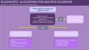
The McDonald criteria, first introduced in 2001, transformed MS diagnosis by formally incorporating MRI into the diagnostic process. Subsequent revisions in 2005, 2010, and 2017 refined this approach, emphasizing MRI and adding CSF oligoclonal bands as supportive markers, enabling earlier and more reliable diagnosis. Traditionally, MS diagnosis required demonstrating dissemination in space (DIS) and dissemination in time (DIT), representing separate demyelinating events across different CNS regions and time points.

However, this long-standing model is now being reconsidered. The 2024 revision introduces a more biologically grounded diagnostic framework that integrates blood and CSF biomarkers, optic nerve assessment via MRI and OCT, and radiological markers such as the central vein sign (CVS) and paramagnetic rim lesions (PRLs). Importantly, the updated criteria also allow certain cases of radiologically isolated syndrome (RIS) to be classified as MS, marking a significant conceptual shift in the diagnostic landscape.

Fig 1: Tips and tricks in applying the 2024 revised McDonald criteria for multiple sclerosis diagnosis.
Fig 2: Clinical presentation and updated approach to radiologically isolated syndrome (RIS) in the 2024 revised McDonald criteria.

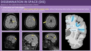
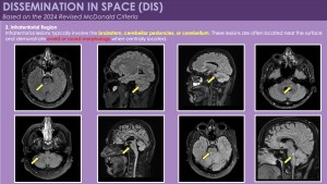
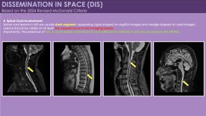
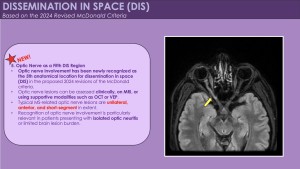
1. Dissemination in Space (DIS): Updated MRI Definition

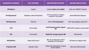
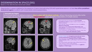
According to the 2024 McDonald criteria, dissemination in space is demonstrated by typical demyelinating lesions in at least two of five anatomical regions:

  • Periventricular
  • Juxtacortical / Cortical
  • Infratentorial
  • Spinal cord
  • Optic nerve (newly included)

The inclusion of the optic nerve represents a major imaging update and supports earlier diagnosis, particularly in patients presenting with optic neuritis. Optic nerve involvement can be confirmed using orbital MRI, supported by OCT and visual evoked potentials (VEP) when required.

Fig 3: Dissemination in space (DIS) according to the 2024 revised McDonald criteria. DIS is defined by typical demyelinating T2/FLAIR hyperintense lesions in at least two of five anatomical regions. The figure demonstrates characteristic periventricular involvement, including lesions in direct contact with the lateral ventricles and Dawson’s fingers, which represent hallmark imaging features of multiple sclerosis.
Fig 4: Juxtacortical and cortical involvement in dissemination in space (DIS). Juxtacortical lesions are defined by direct contact with the cerebral cortex without intervening normal white matter. The schematic illustration on the right highlights common cortical and juxtacortical lesion shapes, including ovoid, U-shaped, and triangular patterns, which are typically observed in multiple sclerosis.
Fig 5: Infratentorial involvement in dissemination in space (DIS). Infratentorial lesions typically involve the brainstem, cerebellar peduncles, or cerebellum. These lesions are often located near the surface and commonly show an ovoid or round morphology on T2/FLAIR images, supporting the assessment of DIS according to the 2024 revised McDonald criteria.
Fig 6: Spinal cord involvement in dissemination in space (DIS). Spinal cord lesions in multiple sclerosis are typically short-segment lesions, appearing cigar-shaped on sagittal images and wedge-shaped on axial images. Lesions should be confirmed on at least two sequences or in two imaging planes. According to the 2024 revised McDonald criteria, the presence of two or more spinal cord lesions is sufficient to fulfill DIS in primary progressive multiple sclerosis.
Fig 7: Optic nerve involvement as the fifth region for dissemination in space (DIS). The 2024 revised McDonald criteria newly recognise the optic nerve as a fifth anatomical region for DIS. Optic nerve involvement may be assessed clinically or on MRI, with supportive modalities such as OCT or VEP. Typical MS-related optic nerve lesions are unilateral, anterior, and short-segment, facilitating earlier diagnosis in patients presenting with isolated optic neuritis.

2. Dissemination in Time (DIT): Flexible Imaging Requirements

MRI-based evidence of DIT may still be demonstrated by:

  •       Simultaneous presence of gadolinium-enhancing and non-enhancing lesions on a single MRI
  •       Appearance of new T2 or enhancing lesions on follow-up imaging

However, under the 2024 criteria, DIT is no longer mandatory when robust biological or topographic evidence is present. This change reduces dependence on longitudinal imaging and allows earlier diagnosis.

Fig 8: Dissemination in time (DIT) according to the 2024 revised McDonald criteria. DIT may be demonstrated by the appearance of new lesions on follow-up MRI, the coexistence of gadolinium-enhancing and non-enhancing lesions, or the presence of positive CSF biomarkers, including oligoclonal bands or an elevated kappa free light chain index. Importantly, DIT is no longer mandatory when dissemination in space involves four or more of the five predefined regions.

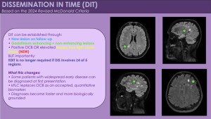
3. MRI-Only Diagnosis in Widespread Disease

In patients with typical clinical presentations, MS can be diagnosed based on imaging alone when:

  •       Lesions are present in ≥4 of the 5 DIS regions

In this scenario, neither DIT nor CSF biomarkers are required, provided that lesion morphology and distribution are characteristic of MS.

Fig 9: MRI biomarkers supporting multiple sclerosis diagnosis according to the 2024 revised McDonald criteria. Advanced MRI biomarkers, particularly the central vein sign (CVS) and paramagnetic rim lesions (PRLs), provide in vivo markers of perivenular and chronic active inflammation, respectively. These imaging features enhance diagnostic specificity and support MS diagnosis when integrated with clinical presentation and lesion topography.

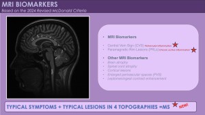
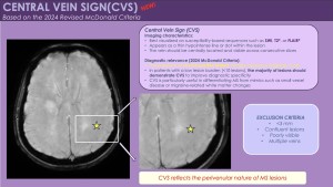
4. Central Vein Sign (CVS): Imaging Marker of Specificity

The central vein sign (CVS) is a radiological biomarker that increases diagnostic specificity for MS:

  •       Defined as a small vein running through the centre of a white matter lesion
  •       Best visualized on SWI, T2*, or FLAIR* sequences
  •       ≥6 CVS-positive lesions strongly support MS diagnosis

In patients with fewer than 10 white matter lesions, the majority of lesions should demonstrate CVS to maintain specificity.

Fig 10: Central vein sign (CVS) as an MRI biomarker in multiple sclerosis. The central vein sign appears as a thin hypointense line or dot centrally located within a white matter lesion on susceptibility-based MRI sequences. According to the 2024 revised McDonald criteria, the presence of multiple CVS-positive lesions increases diagnostic specificity for multiple sclerosis and helps differentiate MS from mimicking conditions.

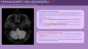
5. Paramagnetic Rim Lesions (PRLs): Chronic Active Lesions

PRLs represent chronically active demyelinating lesions characterised by:

  •       A hypointense rim on susceptibility-weighted imaging
  •       Iron-laden activated microglia at the lesion edge
  •       High specificity (>90%) for MS

The presence of even a single PRL provides strong diagnostic support, especially in challenging or atypical cases.

Fig 11: Paramagnetic Rim Lesions (PRLs). Axial susceptibility-based MRI image demonstrates a chronic active multiple sclerosis lesion surrounded by a thin, complete hypointense rim (yellow star), consistent with a paramagnetic rim lesion. The rim reflects iron accumulation within activated microglia and macrophages at the lesion edge.

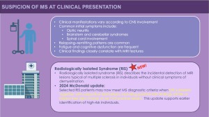
6. Imaging Criteria in Radiologically Isolated Syndrome (RIS)

Under the 2024 revision, RIS may fulfil diagnostic criteria for MS when imaging shows:

  •       DIS according to the 5-region definition and
  •       Supportive imaging or biological markers, such as: ≥6 CVS-positive lesions,positive CSF biomarkers (OCB or kFLC)

This represents a fundamental shift in the imaging-based interpretation of RIS.

7. Imaging in Progressive Multiple Sclerosis

The same imaging framework applies to both relapsing and progressive MS.For primary progressive MS (PPMS):

  •       ≥2 spinal cord T2-hyperintense lesions are sufficient to meet DIS
  •       This reflects the strong predilection of MS for spinal cord involvement in progressive disease

Spinal MRI, therefore, plays a critical role in this subgroup.

8. Recommended MRI Protocols

To reliably detect DIS, DIT, CVS, and PRLs, the following MRI sequences are recommended:

  •       3D FLAIR
  •       T2-weighted imaging
  •       SWI or T2* for CVS and PRLs
  •       Post-contrast 3D T1-weighted imaging
  •       Thin-slice spinal cord MRI (≤3 mm)

Standardised protocols improve reproducibility and diagnostic confidence.

Fig 12: Diagnostic algorithm for multiple sclerosis based on the 2024 Revised McDonald Criteria.
Fig 13: Key differences between the 2017 and 2024 Revised McDonald Criteria

GALLERY