From 2017 to 2023, 7 cases of CFE were diagnosed in our center with an average age of 35.5 years (range: 19-85 years). Of these patients, 6 were male (86%) and 1 was female (14%). All patients were polytraumatized, with fractures of the lower limbs found in all cases at the time of access to the emergency room. In addition, 5 patients (71%) also had fractures of the upper limbs.
At the time of admission, the CT scan of the skull and the CT angiography of the epiaortic vessels were negative for post-traumatic lesions in all 7 patients (100%).
All patients, at an average distance of 4.7 days (range: 1-17 days) from the trauma, developed an altered state of consciousness, requiring urgent CT. The results of urgent CT were negative for lesions in 7 patients (100%).
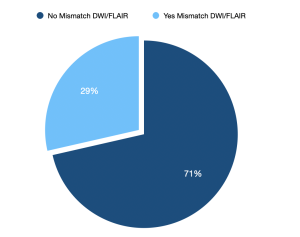
MRI without a contrast agent, confirmed the diagnosis of CFE in all patients. The images showed lesions with proton diffusivity restriction (hyperintensity in DWI, and hypointensity in Apparent Diffusion Coefficient, ADC), areas of vasogenic edema (hyperintensity in T2-FLAIR) and hemorrhagic petechias (hypointensity in Susceptibility-Weighted Imaging, SWI).
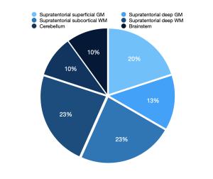
It was observed that the lesions with proton restriction had a ubiquitous distribution both in the supratentorial and infratentorial sites, involving both white and gray matter