The Controlled-Attenuation Parameter (CAP) measured using vibration-controlled transient elastography VCTE (FibroScan®) is a widely used method for quantifying liver steatosis and demonstrates good diagnostic performance.
During Fibroscan evaluation, patients should lie supine with their right hand elevated. The probe is positioned perpendicular to the skin in an intercostal space adjacent to the right lobe of the liver—typically between the 9th and 11th intercostal spaces along the midaxillary line. CAP calculates the attenuation slope of the ultrasound by tracking impulses during the liver stiffness measurement process (Fig. 1).
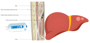
Research indicates that measurements may be unreliable in patients with a skin-liver capsule distance (SCD) exceeding 25 mm. To address this, the XL probe can be used to increase detection depth. Despite these advancements, discrepancies in predicting steatosis grades have been noted even with the XL probe. This suggests that additional factors may influence CAP values. This study hypothesizes that CAP values are influenced not only by the thickness of the skin-liver capsule but also by its overall density. Moreover, it aims to explore whether the fat and muscle components within this region have differing impacts on CAP measurements.
The goal of this study is to investigate the correlation between the thoracic wall components (specifically in the region where FibroScan is performed) and CAP values, as well as to evaluate how these components may serve as predictors of CAP values. The two main characteristics assessed for each component were thickness and CT attenuation, measured in Hounsfield Units (HU). To our knowledge, no prior study has investigated how different skin-liver capsule components might affect CAP values.