
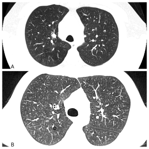
Bronchiolitis or small airways disease refers to injury affecting the bronchioles and alveolar ducts, leading to inflammation or fibrosis. Bronchioles are small airways with a diameter of 2 mm or less located at the center of the secondary pulmonary lobule (SPL). Normally, these small airways are imperceptible on imaging but become visible when abnormal (fig.1).
SPL is the functional unit of the lung and is key to comprehending chest terminology and physiology of multiple diseases. A nuanced knowledge of the distribution of its internal and external structures (fig.2) empowers radiologists to interpret imaging findings accurately.
Bronchiolitis encompasses a broad spectrum of numerous diseases, often presenting nonspecific clinical manifestations that range from an insidious onset of cough and shortness of breath to an acute fulminant illness. The etiology, clinical management and treatment vary among these disorders. Therefore, distinguishing the subtypes of bronchiolitis is crucial for optimizing patients' prognosis.
Small airways diseases are typically not detected in chest radiography. However, chest CT imaging, especially high-resolution CT, almost always reveals abnormal findings, playing a vital role in detecting and classifying small airways diseases. Radiologists are crucial in achieving an accurate diagnosis, improving patient management and prognosis.