
Congress:
ECR24
Poster Number:
C-10866
Type:
EPOS Radiologist (educational)
DOI:
10.26044/ecr2024/C-10866
Authorblock:
J. López Martín, M. M. Merideño García, A. Enriquez Puga, A. A. Gil, E. Ponte, E. F. Berríos, M. S. Paez Alvarez, P. Garcés Marín, A. D. Onuta; Toledo/ES
Disclosures:
Jaime López Martín:
Nothing to disclose
María Montaña Merideño García:
Nothing to disclose
Andres Enriquez Puga:
Nothing to disclose
Asunción Almenar Gil:
Nothing to disclose
Elisabetta Ponte:
Nothing to disclose
Esnelly Francismaría Berríos:
Nothing to disclose
Manuel Sebastian Paez Alvarez:
Nothing to disclose
Pablo Garcés Marín:
Nothing to disclose
Andrei Daniel Onuta:
Nothing to disclose
Keywords:
Lung, Respiratory system, Thorax, CT, Plain radiographic studies, Education, Infection, Inflammation, Transplantation
Bronchiolitis, encompassing a spectrum of airway pathologies with significant clinical impact and varying treatments, poses a challenge for radiologists due to the frequent overlap of imaging findings. These pathologies are categorized into cellular bronchiolitis, characterized by centrilobular nodules with or without a 'tree-in-bud' pattern, and fibrotic bronchiolitis, associated with mosaic attenuation pattern. Within cellular bronchiolitis, numerous entities exhibit distinct imaging findings and associated clinical data that aid in diagnosis. Fibrotic bronchiolitis represents a pattern often associated with a clear clinical background, such as lung transplantation or connective tissue diseases (table 1).
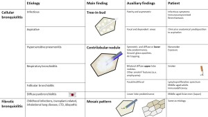
Table 1: Figure 26. Small airways diseases summary, including etiology, pathophysiology, main imaging findings, auxiliary findings, and relevant clinical information.