Congress:
ECR25
Poster Number:
C-19149
Type:
Poster: EPOS Radiologist (educational)
DOI:
10.26044/ecr2025/C-19149
Authorblock:
M. R. Cozcolluela Cabrejas1, J. Gallego1, L. R. Zalazar1, I. Moreno1, A. Perez Del Barrio1, A. Sanz-Cozcolluela2, A. Iturralde1, E. Oliver1; 1Tudela/ES, 2Delft/NL
Disclosures:
Maria Rosa Cozcolluela Cabrejas:
Nothing to disclose
Julio Gallego:
Nothing to disclose
Laura Romina Zalazar:
Nothing to disclose
Irene Moreno:
Nothing to disclose
Amaia Perez Del Barrio:
Nothing to disclose
Ana Sanz-Cozcolluela:
Nothing to disclose
Amaya Iturralde:
Nothing to disclose
Elena Oliver:
Nothing to disclose
Keywords:
Contrast agents, Gastrointestinal tract, Conventional radiography, Fluoroscopy, Barium meal, Complications, Contrast agent-oral, Pathology
RADIOLOGICAL TECHNIQUE
The esophagogram is based on the acquisition of multiple sequential images of the esophagus after the ingestion of effervescent powders and barium contrast.
PATIENT POSITIONING
During the swallowing assessment, a lateral projection along with an anteroposterior (AP) projection, are acquired. Perform left posterior oblique projections in the supine position, where the patient holds a cup with a straw in their left hand (Fig 1).
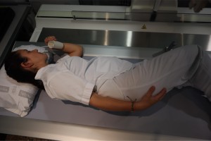
Fig 1: The study begins in a left posterior oblique supine decubitus projection, with the patient holding a cup with their left hand.
To visualize the entry of contrast into the gastric chamber, use a supine/anterior right oblique projection (Fig 2).
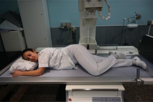
Fig 2: Right anterior oblique/supine decubitus projection. In this position, the gastric antrum, duodenal bulb, and duodenal loop can also be studied.
It is recommended to conclude the study with the patient in an upright position.