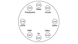
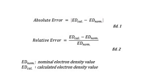
The phantom used in this study was the Catphan600-CTP404 (Phantom Laboratory, NY, USA). As shown in Fig 1, the Catphan600-CTP404 is equipped with eight different modules Air, PMP, LDPE, Water, Polystyrene, Acrylic, Delrin, and Teflon. Each module consists of a cylinder, with an actual diameter of approximately 13 mm and a length of 25 mm along the z-axis. This phantom was scanned using the clinical research CZT-based PCD-CT (TSX-501R, Canon Medical Systems, Otawara, Japan) with the following scanning conditions. Tube voltage: 120 kVp, tube current: 50, 100, 200, 250 mA, rotation time: 0.5 s/rotation, pitch factor: 0.8, CTDIvol: 1.9, 3.9, 7.7, 9.7 mGy, scan mode: helical, focal size: 0.4 mm × 0.5 mm, beam collimation: 0.62 mm × 64. The reconstitution conditions were as follows. Image thickness: 5 mm, image matrix: 512 × 512, reconstruction kernel: hybrid- iterative reconstruction. From the obtained raw data, spectral data based on 5 energies were reconstructed using research software [4]. The electron density values for each of the eight phantom modules were calculated using spectral analysis software on the Vitrea workstation (Canon Medical Systems). The calculated electron density values were compared with the nominal values given in the phantom data sheets, and the absolute and relative errors were calculated according to the following equations Ed.1 and 2. (Fig 2).