Back to the list
Congress: ECR25
Poster Number: C-19654
Type: Poster: EPOS Radiologist (scientific)
Authorblock: M. Shiri1, C. Bortolotto2, A. Bruno1, A. Consonni2, D. M. Grasso2, D. Loiacono1, L. Preda2; 1Milan/IT, 2Pavia/IT
Disclosures:
Mahshid Shiri: Nothing to disclose
Chandra Bortolotto: Nothing to disclose
Alessandro Bruno: Nothing to disclose
Alessio Consonni: Nothing to disclose
Daniela Maria Grasso: Nothing to disclose
Daniele Loiacono: Nothing to disclose
Lorenzo Preda: Nothing to disclose
Keywords: Artificial Intelligence, Lung, Thorax, CT, Image manipulation / Reconstruction, Neural networks, Computer Applications-3D, Computer Applications-Virtual imaging, Image verification
Purpose

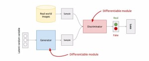
Artificial Intelligence (AI) is increasingly valuable in supporting radiologists, particularly in medical image interpretation, improving diagnostic accuracy, and expediting therapeutic interventions [1]. However, its widespread adoption is still limited by the lack of large annotated datasets, which are challenging to create due to privacy concerns, high costs, and ethical requirements [2, 3]. In light of these challenges, the expansion of datasets with synthetic labeled data is of increasing interest to researchers and clinicians.Generative Adversarial Networks (GANs) [5], offer a promising solution by generating realistic and entirely new medical images that replicate the variability and complexity of the images used in clinical practice. GANs consist of two neural networks, a Generator and a Discriminator, working in a minimax game to improve performance iteratively.

Fig 1: Generative Adversarial Network (GAN) structure.
In the specific area of CT image generation, Ferreira et al. [6] and Han et al. [4] used Progressive Growing (PG)-GAN and Multi-Conditional (MC)-GAN to create lung parenchyma and nodule images. However, these methods are limited to resolutions of 128³ or smaller due to limited memory during training [7]. Memory-efficient GANs aim to address this issue by balancing memory use with generative capability. Lei et al. [8], Yu et al. [9] and  Uzunova et al. [10] proposed slice- or patch-based models or two-step GAN system that, although sparing working memory, generate images with patch artifacts or lack of comprehensive understanding of the entire image structure.

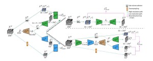
Sun et al. [7] developed HA-GAN, an end-to-end hierarchical architecture generating 256³ resolution images. HA-GAN uses a low-resolution GAN to capture global structure and a high-resolution GAN to refine patches.

Fig 2: HA-GAN structure.
It outperforms models like WGAN [12], VAE-GAN [13], and 3D StyleGAN 2 [14], overcoming memory constraints that limit other models. For this reason, our proposed CRF-GAN is directly compared to HA-GAN.

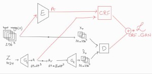
This study addresses the limitations of existing 3D GAN models by proposing CRF-GAN, a novel memory-efficient architecture that combines Conditional Random Fields (CRFs) with GANs to reduce memory usage and improve performance.

Fig 3: CRF-GAN structure.

The key contributions of this work are:

  1. Evaluating CRF-GAN’s clinical value compared to HA-GAN through:(a) Qualitative assessment (primary endpoint).(b) Quantitative assessment (secondary endpoint).
  2. Developing of a memory-efficient GAN architecture using CRFs to enhance anatomical structure consistency in synthetic images.

GALLERY