Congress:
ECR25
Poster Number:
C-16938
Type:
Poster: EPOS Radiologist (scientific)
Authorblock:
M. Woisetschlager1, T. Bjerner1, M. Lindblom1, C. Götz2, A. Hummer2, C. Salzlechner2, A. Spångeus1; 1Linköping/SE, 2Vienna/AT
Disclosures:
Mischa Woisetschlager:
Nothing to disclose
Tomas Bjerner:
Nothing to disclose
Maria Lindblom:
Nothing to disclose
Christoph Götz:
Employee: IB lab GmbH
Allan Hummer:
Employee: IB lab GmbH
Christoph Salzlechner:
Employee: IB lab GmbH
Anna Spångeus:
Advisory Board: UCB Consultant: Giddeon Richter Speaker: Amgen, Tromp Medical
Keywords:
Abdomen, Artificial Intelligence, Computer applications, CT, Computer Applications-General, Osteoporosis
Entire cohort: Compared to the ground truth, the AI demonstrated high accuracy (0.93), sensitivity (0.86), and specificity (0.99) in detecting moderate to severe VFs.
Subgroup analysis: Subgroup analysis indicated accuracy ranging from 0.88 to 0.96, with higher accuracy observed in females compared to males (0.96 vs. 0.89, p=0.03) and in scans using non-bone versus bone kernel protocols (0.96 vs. 0.88, p=0.02). No significant differences were found concerning age, contrast agent use, or anatomic region.
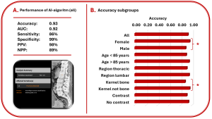
Fig 3: Summary of the results regarding the entire cohort (A) and the sub-groups (B). * indicates a p-value < 0.05.