Cohort: The study utilized 246 existing CT scans originally conducted for purposes unrelated to vertebral assessment but which included the thoracic and/or lumbar spine from a prior study on in-hospital falls. The cohort had a mean age of 84 years (range 62-103) with 42% being female. The CT scans included both women and men, thoracic or abdominal examinations, and were performed with various CT protocols, including bone and non-bone kernel, as well as different contrast phases.
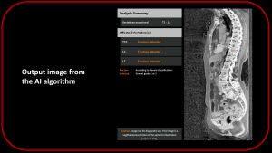
Method: AI analysis was performed using IB Lab FLAMINGO software, which detects vertebral fractures through two AI components: vertebra identification (vertebrae number) and fracture detection (moderate-severe VF, Genant grade 2-3). As ground truth, CT scans were evaluated by two independent and experienced radiologists (>15 years of experience).
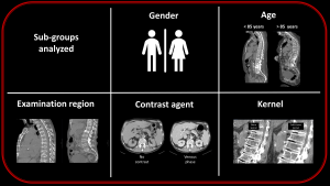
Outcome: The comparison between AI and ground truth was conducted for the entire cohort as well as through subgroup analysis, including sex (male:female), age (<85:>85 years), reconstruction kernel (bone kernel:non-bone kernel), examination region (thorax:abdomen), and use of contrast agent (contrast-enhanced:non-contrast-enhanced).