Congress:
ECR25
Poster Number:
C-20117
Type:
Poster: EPOS Radiographer (scientific)
Authorblock:
P. Clinch, G. Havariyoun, V. Piccio, S. Rees, C. Forster, J. Clinch; London/UK
Disclosures:
Patricia Clinch:
Nothing to disclose
Glafkos Havariyoun:
Nothing to disclose
Vincent Piccio:
Nothing to disclose
Sian Rees:
Nothing to disclose
Charlotte Forster:
Nothing to disclose
James Clinch:
Nothing to disclose
Keywords:
Paediatric, Radiation physics, Radioprotection / Radiation dose, Conventional radiography, Dosimetry, Radiation safety, Dosimetric comparison
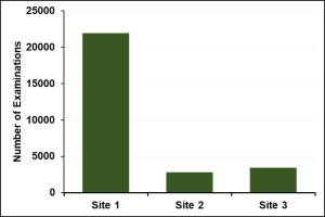
Fig 1: Number of paediatric examinations analysed in the study
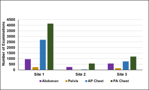
Fig 2: Number and type of paediatric examinations analysed against European DRLs
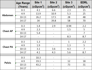
Table 1: Median DAP values from the 3 sites for examinations that can be compared to European DRLs. Missing data denotes lack of data.
From Table 1, the majority of local DRLs for all sites were significantly lower than the European DRLs. The exceptions were;
- Site 1, where the local DRLs for Pelvis examinations (4-9 year old) (n=79) and (10-13 year old) (n=88) exceeded the EDRL by 7.2% and 39% respectively
- Site 2, where the local DRL for Chest examinations (0-3 year old) (n=178) exceeded the EDRL by 32% and
- Site 3, where the local DRL for Pelvis AP examinations (10-13 year old) (n=74), exceeded the European DRL by 135%.
In addition to these local DRLs being established, an additional
- 29 local DRLs were established at Site 1,
- 14 local DRLs were established at Site 2 and
- 32 local DRLs were established at Site 3.
Examples of these local DRLs included Ankle, Clavicle, Femur, Foot, Hand, Knee, Wrist, Hip, Finger, Spine and Elbow.
Where sufficient data was available, median DAPs in rooms where the same examinations were performed were compared at each of the three sites. Differences in DAPs of >20% were seen in most age ranges for chest, elbow, foot, ankle and wrist examinations, when compared to the site median DAP for that examination. Examples are shown in Fig 3 and Fig 4.
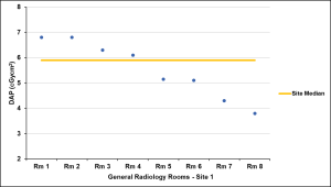
Fig 3: Figure 3: Median DAP values from Site 1 for XANK (10-13 years old) compared with Site median.
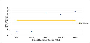
Fig 4: Median DAP values from Site 3 for Elbow examinations (10-13 years old) compared with Site median.