This quantitative study focuses on imaging changes in the supraspinatus muscle tendons. It uses a descriptive-correlational approach to analyze ultrasound data with sample variables. The research was conducted in the Laboratory of Ultrasonography at the School of Health, University of Algarve, Gambelas Campus, Faro.
The study sample consisted of 60 individuals, 36 female individuals and 24 male individuals aged between 18 and 28, grouped into two groups: 28 volleyball players and 32 non-sports practitioners. Data was collected through the application of a questionnaire and an ultrasound examination of both shoulders of the participants. Sampling was non-probabilistic and convenience based. Descriptive and inferential statistics were performed.
Data collection instruments included a high-resolution FUJIFILM ARIETTA 65 ultrasound device with a linear probe and a questionnaire. The questionnaire covered sociodemographic details (age, gender, and occupation) and the participants' volleyball practice and years of practice, including played position, duration, frequency, type of activity, and intensity.
Ethical considerations, including informed consent, confidentiality, and academic integrity, were rigorously adhered to. Formal approvals were obtained from the School Director, the Data Protection Officer, and the University Ethics Committee. Participants provided written consent before data collection began.
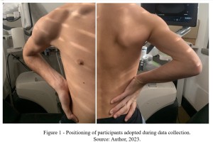
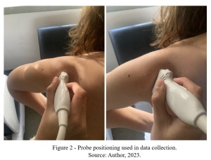
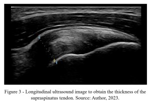
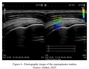
Data collection used ultrasound imaging with participants seated with their elbow flexed at 90º and their hand placed on the iliac crest. The probe was placed in a plane parallel to the one between the collateral shoulder and the ipsilateral hip (Figures 1-2), enabling real-time, two-dimensional imaging (B-mode) and elastography assessment. Measurements were taken three times per tendon to ensure accuracy. The thickness of the right and left supraspinatus muscle tendons was carefully measured using this standardized approach, ensuring comprehensive data collection for comparative analysis, as shown in Figure 3-4 (Panero, et al., 2016; Selame et al., 2021).
Statistical analysis utilized the Statistical Package for the Social Sciences (SPSS) software for descriptive and inferential analyses. Tables and figures were generated to present findings and support comparisons between groups. This methodological approach ensures robust data collection and meaningful interpretation of supraspinatus muscle tendon alterations.