Back to the list
Congress: ECR25
Poster Number: C-27521
Type: Poster: EPOS Radiologist (educational)
DOI: 10.26044/ecr2025/C-27521
Authorblock: S. Khandual, S. Rajan, A. Suman, M. Barnwal; New Delhi/IN
Disclosures:
Swati Khandual: Author: Mahajan Imaging Private Limited
Sriram Rajan: Consultant: Mahajan Imaging Private Limited
Abhishek Suman: Consultant: Mahajan Imaging Private Limited
Madhuri Barnwal: Author: Mahajan Imaging Private Limited
Keywords: Anatomy, CNS, MR-Functional imaging, Comparative studies, Education and training
Findings and procedure details

The scan began with a structural T1-weighted MRI scan. During this anatomical scan, relaxing music was played through a microphone system to keep participants calm and alert, reducing the risk of drowsiness or inattentiveness. Once the anatomical scan was completed, participants were informed that the functional run would begin shortly. Technicians provided verbal instructions to reinforce the task requirements and confirm the participant’s readiness. The experimental task consisted of a 30-second block design, alternating between tapping and rest periods. Ten participants (3 females, 7 males; 9 right-handed, 1 left-handed) performed the task. The task began with unimanual tapping for both the right and left hands, followed by multi-sequential tapping with both hands.

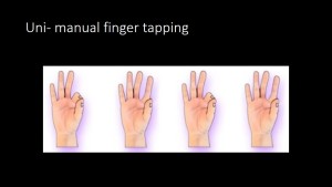
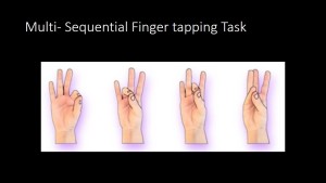
Inside the scanner, participants performed 30 seconds of finger tapping followed by 30 seconds of rest, repeated for three minutes for both hands. The total duration of the paradigm was 8-10 minutes

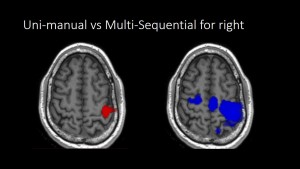
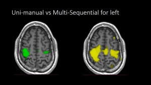
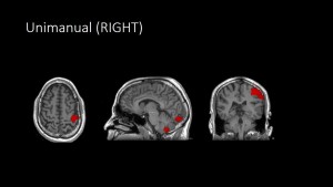
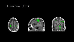
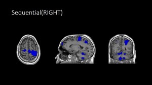
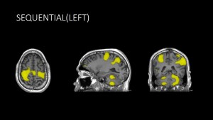
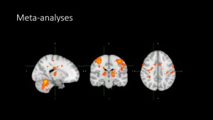
Unimanual finger tapping primarily activated the premotor cortex, specifically the hand region of the motor homunculus, highlighting its specificity and limitations in motor tasks. In contrast, multisequential finger tapping increased the degree of activation of the primary motor area. Additional regions, including the basal ganglia network and cerebellar areas responsible for proprioception & procedural activity were also engaged in the multi-sequential finger tapping, indicating the involvement of more complex cognitive functions. The meta-analysis identified 13 distinct brain regions across cortical, subcortical, and cerebellar areas with significant activity related to tapping tasks. All these findings, validated using thresholds (Z > 4, >100 voxels).

Fig 3: Uni-manual (left sided image, with red activation) vs Multi-sequential (right sided image with blue activation) BOLD activity for right hand. Increased activation of motor cortex seen in the Multi-Sequential activity
Fig 4: Uni-manual (left sided image with green activation) vs Multi-sequential (right sided image with yellow activation) BOLD activity for left hand. Increased activation of motor cortex seen in the Multi-Sequential activity, including of the opposite cortex
Fig 5: Uni-manual finger tapping movement BOLD activations for left hand in whole brain. Mild activation in the occipital cortex and cerebellum also noted.
Fig 6: Uni-manual finger tapping movement BOLD activations for left hand in whole brain. Similar activation of motor cortex, opposite cortex, occipital and cerebellar regions.
Fig 7: Multi-Sequential finger tapping movement BOLD activations for right hand in whole brain. Larger activation of the motor cortex and cerebellar activation
Fig 8: Multi-Sequential finger tapping movement BOLD activations for left hand in whole brain.Larger activation of the motor cortex and cerebellar activation
Fig 9: Merged BOLD activity for multi sequential finger tapping from meta-analyses of 75 studies in Neurosynth. Merged images showing consistent activation in the motor cortex, basal ganglia and cerebellum.
[fig]

GALLERY