Congress:
ECR25
Poster Number:
C-11365
Type:
Poster: EPOS Radiologist (educational)
DOI:
10.26044/ecr2025/C-11365
Authorblock:
P. Del Nido Recio, A. Paternain Nuin, C. Urtasun Iriarte, M. B. Barrio Piqueras, M. Jiménez Vázquez, M. R. López De La Torre Carretero, C. Mbongo, D. A. Zambrano, J. D. Aquerreta; Pamplona/ES
Disclosures:
Pablo Del Nido Recio:
Nothing to disclose
Alberto Paternain Nuin:
Nothing to disclose
Cesar Urtasun Iriarte:
Nothing to disclose
Miguel Barrio Barrio Piqueras:
Nothing to disclose
Marcos Jiménez Vázquez:
Nothing to disclose
Manuel Rafael López De La Torre Carretero:
Nothing to disclose
Carmen Mbongo:
Nothing to disclose
Daniel Alfonso Zambrano:
Nothing to disclose
Jesús Dámaso Aquerreta:
Nothing to disclose
Keywords:
Musculoskeletal soft tissue, MR-Diffusion/Perfusion, Diagnostic procedure, Cancer
Fibroblastic and myofibroblastic tumors can be classified according to their biologic behavior (figure 2).
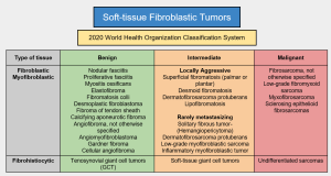
- Benign lesions:
- Elastofibroma dorsi (figure 3): it appears at the scapulo-thoracic joint and it may be heterogeneous on imaging (fibrous and fatty tissue).

- Myositis Ossificans (figure 4): it shows a heterogeneous appearance on imaging and has calcified areas.

- Fibromatosis Colli (figure 5): it is found on the sternocleidomastoid muscle and it is secondary to trauma during childbirth.

- Elastofibroma dorsi (figure 3): it appears at the scapulo-thoracic joint and it may be heterogeneous on imaging (fibrous and fatty tissue).
- Intermediate lesions (locally aggressive or rarely metastasizing) (figures 6-22): Palmar (Dyupuytren) and Plantar (Ledderhose) fibromatosis (figures 11-17) and dermatofibrosarcoma (figures 18-20).


- Malignant lesions (figures 23-27):
- Fibrosarcoma (not otherwise specified): it is hypointense on T1 WI and iso-to hyperintense on T2 WI, with peripheral enhancement after contrast administratio.
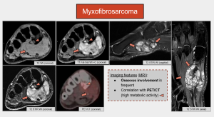
- Myxofibrosarcoma: it has a heterogeneous appearance with hyperintense foci on T2. Osseous involvement is frequent.
- Other tumors that are reviewed are undifferentiated pleomorphic sarcoma, sclerosing epitheloid fibrosarcoma (figures 28 and 29), or low-grade fibromyxoid sarcoma.
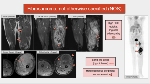
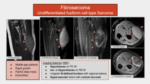
Fibrosarcoma, not otherwise specified (NOS) (figures 23 and 24):
- This tumor has replaced adult fibrosarcoma in the 2020 WHO classification.
- The median patient age at presentation is 50 years.
- It arises from the tendons and fascia of deep soft tissues in the extremities, head and neck, and trunk.
- Histopathology: fibroblast fascicles with a “herringbone” architecture and variable collagen content. Although these features may resemble that of infantile fibrosarcoma, fibrosarcoma NOS has a much worse prognosis.
- CT: isodense infiltrative or well-defined soft-tissue masses.
- MRI: Hypo- to isointense on T1 WI and heterogeneously hyperintense on T2 WI, band-likes areas may be seen (fibrous tissue, hypointense on T1 and T2 WI) and variable enhancement (predominantly peripheral).
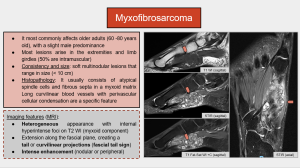
Myxofibrosarcoma (figures 26 and 27):
- It most commonly affects older adults (60 -80 years old), with a slight male predominance.
- Most lesions arise in the extremities and limb girdles (50% are intramuscular).
- Consistency and size: soft multinodular lesions that range in size (< 10 cm).
- Histopathology: It usually consists of atypical spindle cells and fibrous septa in a myxoid matrix Long curvilinear blood vessels with perivascular cellular condensation are a specific feature.
- MRI: Heterogeneous appearance with internal hyperintense foci on T2 WI (myxoid component), fascial tail sign and intense enhacement (nodular or peripheral).
Sclerosing Epithelioid Fibrosarcoma (SEF) (figures 28 and 29):
- It usually occurs in middle-aged patients (mean age of 47 years).
- SEF commonly affects lower extremities, pelvic girdle or upper extremities.
- 80% of these tumors metastasize (to the lungs, pleura, bones and brain).
- Histopathology: polygonal epithelioid fibroblasts arranged in cords in a dense, hyalinized, and sclerotic stroma.
-
MRI:
- Variable SI on T1 WI and heterogeneous high SI on T2 WI.
- Necrosis and peripheral enhancement. Enhancement indicates areas of cellularity, vascular proliferation, and peritumoral desmoplastic reaction.