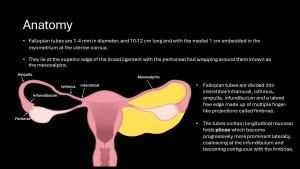
Knowledge of the embryological development and normal anatomy of the fallopian tubes is key to understanding fallopian tube disease.


Imaging
The imaging modality of choice for investigating fallopian tube disease will depend on the presentation. Signs and symptoms can, however, be non-specific and mimic uterine, ovarian, or bowel pathology. These usually include lower abdominal pain, per vaginal discharge or bleeding, and infertility.
Transvaginal ultrasound (TVUS) is usually the first-line investigation. Magnetic resonance imaging (MRI), with higher tissue resolution, is reserved for problem-solving in more complex and atypical cases, and hysterosalpingography (HSG) is used to evaluate tubal patency. However, due to the widespread use of computed tomography (CT) in the emergency setting, fallopian tube pathology may often be detected first on a CT scan performed for other indications.