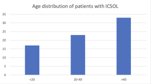
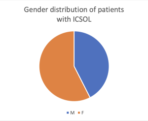
This prospective observational study was conducted at Bharati Hospital and Research Centre, Pune, in the Department of Radiodiagnosis, focusing on patients with suspicion or presence of an intracranial space-occupying lesion (ICSOL).
73 participants were included in the study which was done over 18 months to ensure a diverse range of cases. The sample size was determined through the prevalence formula (p=0.3, q=0.7, d=0.1072).


Patients diagnosed with ICSOLs on CT or MRI or those returning for follow-up after radiotherapy or surgery for residual or recurrent tumors were included in the study. Patients with intraparenchymal hematomas, contraindications to MRI, or inability to cooperate during procedures were excluded from the study. Sampling involved recruitment of patients meeting the criteria, with informed consent obtained from adults and assent secured for children above 7 years, alongside guardian consent.
Data was collected using a custom proforma documenting clinical features, demographics, imaging findings, and histopathological results when relevant. Imaging was performed on a Philips Ingenia Elition 3 Tesla MRI system using multiparametric sequences, including APT, as part of the standard tumor protocol. APTw images were acquired with specific parameters: saturation pulse duration of 2 seconds, frequency offsets from -3.5 to +3.5 ppm, and scan time of 3 minutes and 46 seconds. Imaging parameters included a SENSE factor of 1.6, TR of 1000 ms, TE of 8.3 ms, matrix size of 128 × 128 (reconstructed to 256 × 256), FOV of 230 × 230 mm, and voxel size of 1.8 mm (reconstructed voxel 0.9 mm).
Analysis involved mean ROI values and color maps from APT sequences in combination with conventional MRI sequences (T1, T2, DWI, FLAIR, contrast imaging). ROIs (≤15 mm²) were drawn on areas of maximum contrast enhancement or lowest ADC for non-enhancing lesions, avoiding necrosis, calcifications, or cystic changes. A rainbow color scale (-5% to 5%) facilitated interpretation, with normal brain tissue appearing green and pathological entities displaying varied signals. Data were analyzed using SPSS version 28.0, with results presented as tables and graphs.
Ethical approval was obtained, and the study concluded upon achieving histopathological or clinical diagnoses of ICSOLs. This methodology enabled systematic evaluation of APT imaging as a complementary tool in the characterization and diagnosis of ICSOLs.