Congress:
ECR25
Poster Number:
C-21700
Type:
Poster: EPOS Radiologist (scientific)
Authorblock:
A. Choux1, Z. Yin2, C. L. Kim2, A. Pourmorteza1; 1Atlanta, GA, GA/US, 2Niskayuna, NY/US
Disclosures:
Arnaud Choux:
Research/Grant Support: GE HealthCare
Zhye Yin:
Employee: GE HealthCare
Chang Lyong Kim:
Employee: GE HealthCare
Amir Pourmorteza:
Grant Recipient: GE HealthCare
Keywords:
Computer applications, CT, CT-Angiography, CT-Quantitative, Physics, Cancer
A cylindrical test object (diameter=20cm, height=5cm) was filled with ballistic gel and contained 137 2-mL test tubes filled with calibrated concentrations of materials including: iodine-, gadolinium, tantalum, bismuth-based contrast agents and their mixtures and dilutions in water and simulated blood.
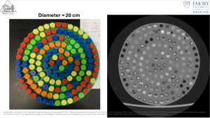
Fig 2: Test object used in the study. Left: Photo. Right: CT reconstruction.
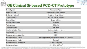
Table 1: Image acquisition and reconstruction parameters.