Congress:
ECR25
Poster Number:
C-27813
Type:
Poster: EPOS Radiologist (scientific)
DOI:
10.26044/ecr2025/C-27813
Authorblock:
S. El Graini, S. Cherkaoui, H. Retal, I. Dokal, Y. Omor, R. Latib, S. Amalik; Rabat/MA
Disclosures:
Soumya El Graini:
Nothing to disclose
Sara Cherkaoui:
Nothing to disclose
Hamza Retal:
Nothing to disclose
Ibrahima Dokal:
Nothing to disclose
Youssef Omor:
Nothing to disclose
Rachida Latib:
Nothing to disclose
Sanae Amalik:
Nothing to disclose
Keywords:
Oncology, MR, Diagnostic procedure, Metastases
This retrospective descriptive study was conducted from January 2022 to December 2024, focusing on patients with brain metastases (BM) from NSCLC, presented with various characteristics and locations. Morphological imaging was used to assess the metastases' number, location, size, and shape. When advanced imaging sequences were available, they allowed for the evaluation of tumor cellularity (diffusion sequence), tumor vascularization (PWI), and metabolic profiles (MRS).
A. CT Scan Protocol
- Patient Positioning: Supine, arms alongside the body.
- Scout View: From the vertex to C2.
- Slice Thickness: <1 mm.
- Contrast Administration:
- IV Contrast Agent: 80–100 cc.
- Injection Rate: 3–5 cc/s.
- Delayed Acquisition: 3–5 minutes post-injection.
- Reconstruction: Multiplanar reconstructions (MPR) in axial, sagittal, and coronal planes with parenchymal and bone window settings.
B. MRI Protocol
- Conventional Sequences
- At least two orthogonal planes: axial and sagittal.
- T1-weighted 3D: Findings: Melanin hypersignal, hemorrhagic metastases.
- Susceptibility-Weighted Imaging (SWI).
- Optional:
- Axial T2-weighted imaging (variable signal).
- High-resolution T2-weighted sequence for cranial nerve pathology assessment.
- Spectroscopy MRS
- Post-Contrast Sequences (Gadolinium Injection)
- Perfusion T2*
- FLAIR: Detection of perilesional edema and leptomeningeal metastases.
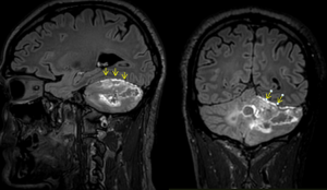
Fig 1: Sagittal and Axial FLAIR sequences after Gadolinium injection reveal well-defined leptomeningeal enhancement, indicative of carcinomatous metastatic disease in a 70-year-old patient with lung adenocarcinoma. This pattern of enhancement is characteristic of leptomeningeal carcinomatosis, highlighting the spread of metastatic cells to the meninges. The FLAIR sequences, combined with contrast, provide valuable information for detecting subtle abnormalities in the brain's lining, aiding in the diagnosis of metastatic involvement in such cases.
- T1-weighted Turbo Spin Echo (TSE):
- Acquired in three planes post-contrast.
- Standard Acquisition: 5 minutes after injection.
- Optional Delayed Acquisition: 75 minutes after injection.