Congress:
ECR24
Poster Number:
C-11913
Type:
EPOS Radiologist (scientific)
Authorblock:
E. Cho, H. J. Baek, Y. Jeong; Changwon/KR
Disclosures:
Eun Cho:
Nothing to disclose
Hye Jin Baek:
Nothing to disclose
Yujin Jeong:
Nothing to disclose
Keywords:
Head and neck, CT, Ultrasound, Biopsy, Cancer, Metastases
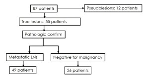
87 patients with suspicious ScLNs on chest CT
- 49 patients (56.3%) : metastatic LN
- 26 patients (29.9%) : negative for malignancy
- 12 patients (13.8%) : pseudo-lesion
- partial volume artifact of the anterior scalene muscles
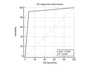
The diagnostic indices of subsequent neck US for the 87 patients with true LNs detected on chest CT
- Sensitivity: 91.8%
- Specificity: 92.3%
- PPV: 95.7%
- NPV: 85.7%
- Accuracy: 92.0%

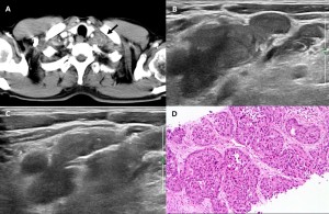
Fig 4: A 64-year-old male with left supraclavicular LN that confirmed as metastasis by US-CNB.
(A) On axial CT scan, there were multiple, marginal enhancing LNs with intranodal non-enhancing portions (arrow) in the left supraclavicular region. (B,C) In corresponding region, there were multiple enlarged L/Ns with loss of normal fatty hilum and conglomerative features, and then US-guided CNB was performed. (D) Epithelioid columnar cells are arranged in complex and cribriform pattern with distinct nuclear atypia and pleomorphism, highly suggestive of metastatic adenocarcinoma. (H&E, x200)
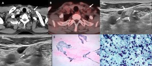
Fig 5: A 78-year-old male with left supraclavicular LN that confirmed as reactive LN by US-FNA.
(A) On axial CT scan, there was small LN (arrow) in the left supraclavicular area. (B) On PEC-CT, there was no significant uptake of the lesion (arrow). (C,D) On US, the corresponding LN showed normal fatty hilum without cortical thickening, suggesting reactive hyperplasia, and then US-FNA was done. (E, F) The aspiration cytology of LN shows many scattered cluster of cells with low cellularity. There are polymorphous benign-looking lymphoid cells without cytological atypia (x400).
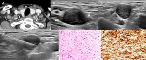
Fig 6: A 49-year-old female with right supraclavicular LN that confirmed as neurogenic tumor by US-CNB.
(A) On axial CT scan, there was a well-defined, mildly enhancing nodule in the right supraclavicular area (dashed circle). ar area. (B-D) On US, the lesion showed a well-defined oval hypoechoic nodule with direct connection to adjacent linear hypoechoic structures (arrows), suggesting neurogenic tumor. Then, US-CNB was performed (E) The pleomorphic spindle cells have short plump nuclei with eosinophilic cytoplasm (H&E, x200). (F) The spindle cells are strong and diffuse positive for S100p, suggestive of neurogenic origin ( x200).

Fig 7: A 71-year-old male with asymmetric anterior scalene muscle, misinterpretation as supraclavicular LN on CT
(A) On axial CT scan, there was a nodular lesion with mild perilesional fat strandings in the left supraclavicular region (dashed circle). (B) On US, there was no pathologic LN in the corresponding area.) (C) On coronal CT scan, the lesion was the anterior scalene muscle.