
Study Population
A retrospective review was performed on 183 patients who underwent NET for intradural aneurysms between May 2019 and February 2023. A total of 194 aneurysms were treated.
All procedures were conducted under institutional review board approval (No. 1976), and informed consent was obtained.
Endovascular Procedure
All NET procedures were conducted using a biplane flat-panel detector system (Azurion7 B20/15; Philips Healthcare). The lateral X-ray tube was positioned on the patient’s left side. Tube voltage and current were automatically modulated by the automatic brightness control system in both fluoroscopy and DSA modes.
The standard workflow consisted of:
- Initial diagnostic DSA (arterial & venous phases)
- Three-dimentional rotational angiography
- Working-projection DSA series during coiling or stent deployment
- Final DSA confirmation
- Cone-beam CT
These steps collectively contribute to cumulative patient skin dose, with angulation-dependent variations influencing dose localization.
Radiation Dosimetry
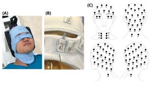
Skin dose mapping was performed using 64 radiophotoluminescence dosimeter (RPLD) chips (GD-302M) integrated into a stretchable polyester dosimetry cap. Each RPLD chip was cylindrical in shape, with a length of 12 mm and a diameter of 1.5 mm. The characteristics of the RPLD and the dose measurement system are as follows (figure 3 and figure 4):
- RPLD demonstrate excellent dose and dose-rate agreement with ionization chamber dosimeters.
- RPLD chip is radiopaque, so it does not interfere with the procedure.
- Chips were arranged to cover the entire scalp surface.
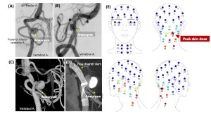
- The highest measured value among the 64 points was defined as the PSD.
- Precise information on entrance skin dose and dose distribution can be obtained.
Data Collection
Patient demographics, aneurysm characteristics, and procedural details were retrospectively reviewed. Collected data were as follows:
- Patient demographics: age, sex and BMI
- Aneurysm characteristics: location (anterior vs. posterior circulation), reccurence and number (single vs. multiple)
- Procedural factors: stent use, total fluoroscopy time (TFT), air kerma at the patient entrance reference point (Ka,r) and kerma-area product (PKA)
- "Ka,r ratio" was defined as the ratio of the Ka,r values of the frontal X-ray tube to the Ka,r value of the lateral X-ray tube.
Statistical Analysis
All analyses were performed using JMP® Pro 18.2.0. Continuous variables are reported as mean ± SD. Logistic regression (univariate and multivariate) was used to identify predictors of occipital PSD, which was the most common location.