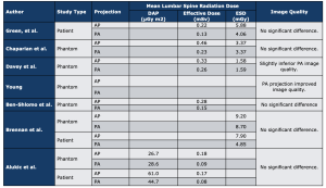
A total of seven studies were included in the qualitative synthesis (Fig. 3). These studies all met the search (inclusion/exclusion) criteria and directly compared lumbar spine radiography performed in the AP and PA positions. In total studies included 210 patients from both clinical and phantom experiments. All studies reported a lower effective dose for the PA projection when compared to the AP. Studies also agreed by consensus that there was no significant loss of image quality or diagnotic acceptability when imaging in a PA projection.
All five of the studies that included effective dose in its analysis found a reduction in dose in the PA position when compared to the AP position.
- Mean reduction in effective dose was 46%.7-11
Possible mechanisms for this reduction include:
- Protection of the more radiosensitive organs
- Tissue redistribution
Although it is clear there is a dose reduction effect on the patient in the PA position for lumbar spine radiographs it is important to take into consideration patient safety and comfort when adopting this technique. In terms of image quality all of the studies included in this review agreed that there was no significant reduction in image quality or diagnostic acceptability in the PA position compared to the AP position.