Acute chest pain is a common and significant health concern, accounting for a substantial number of morbidity and mortality worldwide.
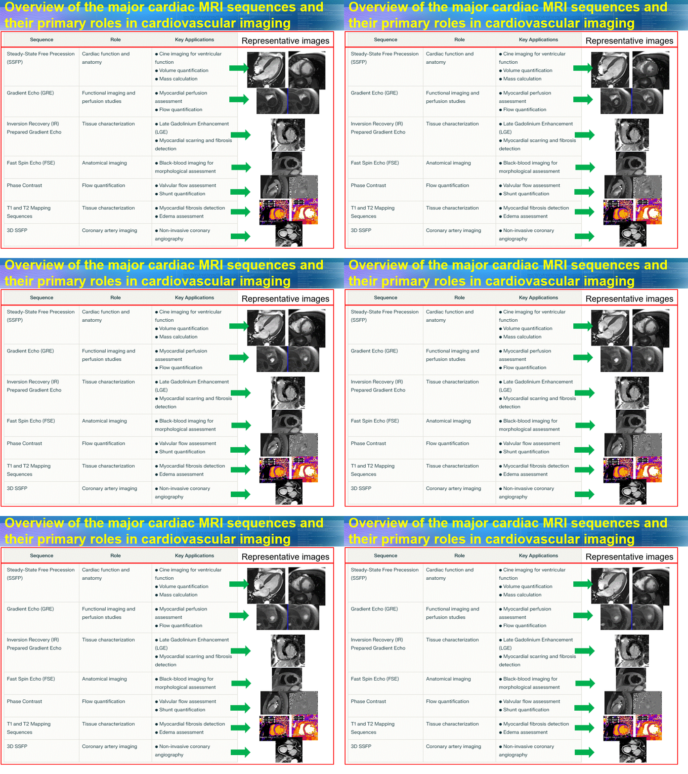
Cardiovascular magnetic resonance (CMR) imaging has evolved significantly over the past three decades, becoming an invaluable tool in cardiac diagnostics. CMR employs several key pulse sequences, each serving specific roles in evaluating cardiovascular structure and function.

The development of MR cardiac stress imaging (MRCSI) has further enhanced its utility, particularly in evaluating patients with suspected coronary artery disease (CAD).
MRCSI offers significant advantages in terms of diagnostic accuracy, comprehensive evaluation, and risk stratification without radiation exposure.
Despite its potential, MRCSI faces several challenges, such as motion artifacts, lengthy scan times, and complex protocols. These issues are exacerbated by the heart’s continuous motion and respiratory cycles, which can lead to image artifacts if not properly managed.
State-of-the-Art Techniques
Recent advancements in MRCSI focus on overcoming these challenges through motion correction and fast imaging sequences. Motion correction techniques, like ECG gating and navigator echo respiratory gating, help mitigate motion artifacts by synchronizing image acquisition with the cardiac cycle1. Fast imaging sequences, such as real-time MRI and compressed sensing, reduce scan times and improve image quality by allowing rapid data acquisition.
Protocol Optimization
Optimizing MRCSI protocols involves selecting appropriate pharmacological agents, such as adenosine or regadenoson, to induce hyperemia. Gadolinium-based contrast agents are used for myocardial perfusion assessment, with precise timing of image acquisition being crucial for accurate results. Additionally, arrhythmia rejection techniques help manage irregular heartbeats by excluding data acquired during arrhythmic events.