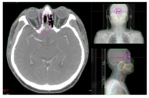
Ten patients with Nasal Cavity Cancer who had undergone Intensity-Modulated Radiation Therapy (IMRT) were randomly selected. Each patient was immobilized using a thermoplastic mask to stabilize the head and neck. CT images with a slice thickness of 2.5mm were obtained from the skull to the clavicles.
For target delineation, the Planning Target Volume (PTV) was defined following the identification of the Gross Tumor Volume (GTV) using preoperative PET-CT and CT scans, adhering to NCCN guidelines. PTVs were delineated to avoid overlap with both eyes.
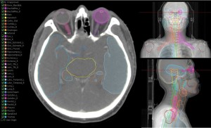
Organs at risk (OARs) were delineated using AI contouring software (Oncosoft), with subsequent manual adjustments. OARs included both eyes, optic nerves, optic chiasm, pituitary gland, brainstem, and lenses.
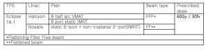
Treatment plans were generated using Eclipse16 RTP software for both Novalis TX's IMRT (static 6-port + non-coplanar 2-port) and Halcyon's IMRT (static 8-port) and Volumetric Arc Therapy (VAMT) (half 8-arc). A comparative dosimetric analysis was performed[3].

Quality assurance of the three plans was conducted using the Delta 4 phantom to verify the pass rates. Parameters including Conformity Index (CI), Heterogeneity Index (HI), 95% target coverage, as well as Dmax and Dmean values of each OAR, were evaluated.