Pre-operative CTA images are an essential part of EVAR planning, offering detailed insights into the morphology of the aneurysm and its surrounding structures. These images are used to measure parameters such as the diameter of the aneurysmal sac, the aortic-iliac diameters, and the presence of critical features like wall thrombus and calcifications. Accurately measuring these features is vital for selecting the correct stent graft size, which directly impacts the procedure’s success. Inaccurate measurements, whether due to human error or variability between different clinicians, can lead to suboptimal planning, resulting in complications such as graft migration, endoleaks, or the need for reintervention.
In this study, we obtained a set of 15 pre-operative CTA scans from patients with AAA, sourced from various open-access medical imaging databases. These scans were used to extract the relevant morphological features needed for EVAR planning. Two experienced vascular surgeons manually segmented and measured the images using dedicated clinical software, while the same features were automatically extracted by the newly developed AI tool.
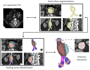
The AI-based tool utilizes advanced 3D image segmentation techniques, leveraging deep learning algorithms to accurately delineate the aortic and iliac vessels and identify key morphological features. The tool’s output is designed to be compatible with clinical workflows, offering automated measurements that can be seamlessly integrated into the pre-operative planning process. The entire pipeline of the AI tool is illustrated in Fig 1, showing the sequence of steps from the input of pre-operative CTA images to the final output of the morphological measurements and features. The pipeline includes image segmentation of the aortic-iliac vessels, sealing zones recognition and extraction of key morphological features compatible with the clinical workflow.
To evaluate the accuracy of the AI tool, we compared the manually extracted measurements to the automatic ones using Bland-Altman plots. Bland-Altman analysis is commonly used to assess agreement between two different measurement methods, and the lack of significant bias between the manual and automatic measurements would demonstrate that the AI tool is capable of replicating the accuracy of experienced clinicians. For categorical data, such as the percentage of wall thrombus or calcification present in the proximal and distal sealing zones, we used Cohen’s Kappa coefficient to assess the level of agreement between the manual and automatic methods. Cohen’s Kappa is a statistical measure of inter-rater agreement for categorical variables, and high Kappa values would indicate that the AI tool performs similarly to the experienced surgeons.
Furthermore, the time taken to extract the necessary morphological features was measured for both methods, with the aim of quantifying any time savings that the AI-based tool could provide. Statistical analysis was performed using the Mann-Whitney U test to compare the extraction times for the manual and automated approaches.