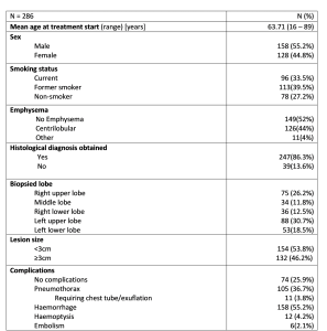
Among 286 included patients, 55% were male. The ages ranged from 16 – 89 years and the average age was 63.71 years. Most of the patients were either former (n=113; 39.5%) or current smokers (n=96 ;33.5%). Definitive pathohistological diagnosis was observed in 247 cases (86.3%). In terms of lesion size 46.2% (n=132) were greater then 3cm while 53.8 (n=154) had a smaller diameter then 3 cm. Complications were observed in 212 (74.1%) patients. The most common complication was hemorrhage (55.2%; n=158), followed by pneumothorax (36.7%; n=105). Only 3.8% (n=11) of evidenced pneumothoracies required intervention while the rest were overt and resolved spontaneously without any worsening of clinical situation.Hemoptysis occurred in 4.2% (n=12) of patients. No cases of massive hemoptysis were observed, and all instances of recorded hemoptysis were transient. In cases of massive parenchymal hemorrhage and hemoptysis patients were administered 1 ampule of tranexamic acid in bolus, after that if needed one more was given trough infusion. No patients that experienced hemorrhage required blood transfusions, embolization or surgical exploration.Embolism was evidenced in 2.1% (n=6) of cases. Two patients experienced gas embolism in brain vascular structures.
The factors that could have led to complication rates such as age (>70 years), sex, smoking status, emphysematous changes and lesion size were evaluated. The mentioned factors were not found to be significantly associated with hemorrhage, embolization or hemoptysis. However, emphysematous changes (p>0.001) and active smoking status (p=0.01) were found to be significantly associated with the risk of pneumothorax development. Occurrence of complications in general (p=0.67), including pneumothorax specifically (p=0.91), was not determined to be a risk factor for the inability to obtain a definitive pathohistological diagnosis.
