Back to the list
Congress: ECR25
Poster Number: C-17410
Type: Poster: EPOS Radiologist (educational)
Authorblock: P-S. Muntean1, D. R. R. Ban2, M. Racheriu2, I-O. Nastase3, M. Sandu2, F. Grosu2; 1Alba Iulia/RO, 2Sibiu/RO, 3Cisnadie/RO
Disclosures:
Paula-Stefana Muntean: Author: Collecting information and composing content.
Delia Rodica Rodica Ban: Consultant: Consultation on the text.
Mihaela Racheriu: Consultant: Granting access to the hospital database, outlining key topics, and reviewing the text.
Ionut-Octavian Nastase: Consultant: Consultation on the text.
Mariana Sandu: Consultant: Consultation on the text.
Florin Grosu: Consultant: Granting access to the hospital database, outlining key topics, and reviewing the text.
Keywords: Musculoskeletal bone, Musculoskeletal joint, Musculoskeletal spine, CT, MR, Plain radiographic studies, Complications, Diagnostic procedure, Education, Education and training, Inflammation
Findings and procedure details

Radiographic imaging remains an essential tool in the diagnosis and monitoring of AS, providing clear indications of early inflammatory changes and advanced bony fusion.

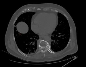
While radiography is crucial in detecting structural changes, CT imaging offers a more detailed and precise visualization of bony structures, allowing for better identification of subtle erosions and ossifications. It is especially useful in evaluating the progression of spinal ankylosis and sacroiliac joint involvement, helping clinicians monitor disease progression and treatment responses.

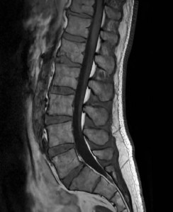
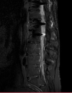
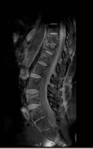
MRI is an invaluable tool for detecting early inflammatory changes in the disease process that may not yet be visible on plain radiographs. In particular, MRI provides superior sensitivity for identifying edema in the vertebral corners and sacroiliac joints and changes in soft tissues, such as ligamentous ossification and disc degeneration. Additionally, MRI is crucial for detecting associated complications, including spinal fractures, spondylodiscitis (Andersson lesions), and sacroiliitis, which can significantly impact treatment strategies.  [2]

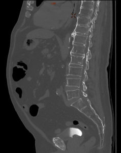
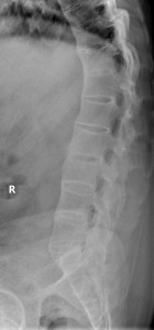
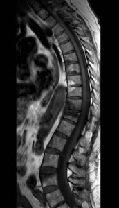
Both radiographically, as well as on CT and MRI, the primary key signs that we can identify in ankylosing spondylitis (AS) at the spinal level are inflammatory changes at the entheses, which progress with edema and erosions at the vertebral corners. In response, osteosclerosis develops at this level, which is radiologically represented as the "Shiny corner sign" or "Romanus lesion", which it is characterized radiographically or on CT by triangular areas at the vertebral corners with an osteosclerotic or osteocondensing appearance , and on MRI by hypointensity on T1 and hyperintensity on STIR (early edematous changes), which later evolve with lipid degeneration, showing hyperintensity on T1. [3][4]

Fig 1: Lumbar spine X-ray - RL view: early changes of ankylosing spondylitis with Romanus lesions at the lower lumbar level
Fig 2: Lumbar spine MRI – Sagital T1: early changes of ankylosing spondylitis with Romanus lesions at the lower lumbar level
Fig 3: Lumbar spine MRI – Sagital STIR: early changes of ankylosing spondylitis with Romanus lesions at the lower lumbar level

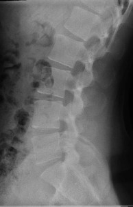
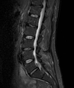
As these changes progress, the concavity of the anterior edges of the vertebral bodies loses its concavity at multiple levels, which is radiologically identified as vertebral body squaring.

Another change that may occur in ankylosing spondylitis as a result of chronic inflammation is the calcification inside a spinal ligament or the annulus fibrosus, which manifests as marginal bony outgrowths with a vertical orientation, known as syndesmophytes.

Fig 4: Lumbar spine X-ray - RL view: specific changes of ankylosing spondylitis with vertebral body squaring, anterior syndesmophytes forming bridges.
Fig 5: Lumbar spine X-ray – AP view: specific changes of ankylosing spondylitis with vertebral body squaring, lateral syndesmophytes forming bridges.
Fig 6: Thoracolumbar spine CT – Sagittal view: specific changes of ankylosing spondylitis with vertebral body squaring, anterior syndesmophytes forming bridges.

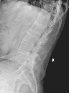
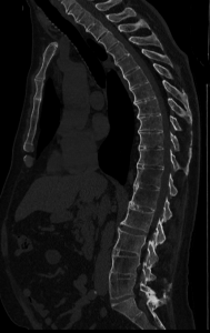
Over time, these can fuse, forming bony blocks which, together with the other described changes, give a radiologic appearance of a "bamboo spine."  This is one of the pathognomonic signs of ankylosing spondylitis.

Fig 7: Lumbar spine X-ray - RL view: specific changes of ankylosing spondylitis with anterior syndesmophytes, ossification ALL, forming bridges, and a 'bamboo spine' appearance. Straightening of the lumbar spine
Fig 8: Lumbar spine MRI – Sagital T1: Narrowing of the intervertebral spaces, ossification of the spinal and interspinous ligaments, and syndesmophytes with a "bamboo spine" appearance, with a tendency for block formation and moderately hyperintense T1 signal of the intervertebral discs; radiosynthesis with bars and transpedicular screws in the thoracolumbar region partially included in the acquisition plane.
Fig 9: Lumbar spine MRI – Sagittal STIR: Narrowing of the intervertebral spaces, ossification of the spinal and interspinous ligaments, and syndesmophytes with a "bamboo spine" appearance, with a tendency for block formation and moderately; radiosynthesis with bars and transpedicular screws in the thoracolumbar region partially included in the acquisition plane.

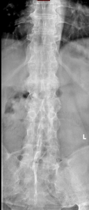
As the disease progresses, a series of spinal ligaments, joints, and discs ossifications may occur. A notable imaging sign visible on radiography, CT, or MRI is the "Dagger sign," characterized by the ossification of the interspinous ligament in the frontal plane. A particular feature of MRI is that enhancement of the interspinous ligaments indicates enthesitis.

Fig 10: Lumbar spine X-ray - AP view: specific changes of ankylosing spondylitis with lateral syndesmophytes, forming bridges and a 'bamboo spine' appearance. The 'Dagger sign' is also visible.

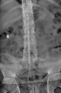
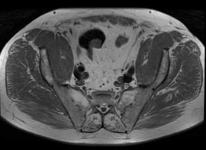
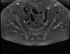
There are also specific imaging changes in AS related to sacroiliac involvement, which sacroiliitis changes can quantify according to the New York or ASAS criteria. This is an essential hallmark of SpA, initially characterized by edematous inflammatory changes that precede structural modifications, visible on MRI (T1 hypointensity, STIR hyperintensity), synovitis, and capsulitis (on gadolinium-enhanced T1). As sacroiliitis progresses, erosions develop, which may initially present as pseudo widening of the joint, detectable on radiographs, CT, and MRI, followed by subchondral osteosclerosis and proliferative changes, ultimately resulting in ankylosis.

Fig 11: X-ray specific to the sacroiliac joint, AP view: changes consistent with grade IV sacroiliitis according to the New York criteria, with narrowing up to the disappearance of the joint space and marginal erosions with ankylosis.
Fig 12: Sacro-iliac joint -para axial T1: Narrowing of the joint space, up to its disappearance at the anterior level, marginal erosions, and small areas of edema and subchondral osteosclerosis.
Fig 13: Sacro-iliac joint -para axial STIR: Narrowing of the joint space, up to its disappearance at the anterior level, marginal erosions, and small areas of edema and subchondral osteosclerosis.

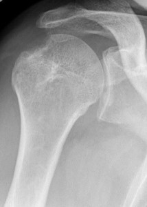
To a lesser extent, AS also affects peripheral joints such as the hip (coxofemoral), knee, shoulder (scapulohumeral), and hand joints, as well as causing various erosions and fusions of the sternoclavicular, costochondral, and costovertebral joints. One of the specific radiological signs of AS affecting the shoulder is the "hatchet" deformity, defined by a well-circumscribed erosion on the lateral-dorsal aspect of the humeral head, resulting in a characteristic hatchet-like shape. [2] [3] [4]

Fig 14: Fig. 14: Right shoulder X-ray – AP view: "hatchet" sign.

From a complication standpoint, several specific radiological signs exist, such as Andersson lesions (called rheumatic spondylodiscitis), as a result of persistent inflammatory changes at the disc-vertebral level, which is characterized radiographically and on CT by irregularities and erosions of the vertebral endplates at the central level, and on MRI by disc-vertebral signal changes, with hyperintensity on STIR and hypointensity on T1 (often hemispherically shaped). [4] [5]

Fig 15: Thoracolumbar spine MRI- Sagital T1: Changes suggestive of ankylosing spondylitis, with the development of syndesmophyte ankylosis, mirror-image edema at the T10-T11 level, associated with disc edema at this level suggestive of an Andersson lesion, secondary to a "chalk stick" fracture, with moderate reduction in vertebral body height and a thin prevertebral fluid layer
Fig 16: Thoracolumbar spine MRI- Sagital T2: Changes suggestive of ankylosing spondylitis, with the development of syndesmophyte ankylosis, mirror-image edema at the T10-T11 level, associated with disc edema at this level suggestive of an Andersson lesion, secondary to a "chalk stick" fracture, with moderate reduction in vertebral body height and a thin prevertebral fluid layer
Fig 17: Thoracolumbar spine MRI- Sagittal STIR: Changes suggestive of ankylosing spondylitis, with the development of syndesmophyte ankylosis, mirror-image edema at the T10-T11 level, associated with disc edema at this level suggestive of an Andersson lesion, secondary to a "chalk stick" fracture, with moderate reduction in vertebral body height and a thin prevertebral fluid layer

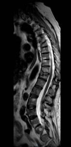
Due to chronic inflammatory status and extensive ossifications, patients with AS are significantly more prone to complications such as fractures, even in cases of minor trauma. One particularly specific type of fracture is the "chalk stick" or "carrot stick" fracture, which commonly occurs in the lower cervical or upper thoracic spine. [4] [5]

Fig 18: Thoracolumbar spine CT – Axial view: 'Chalk stick' fracture of T10
Fig 19: Thoracolumbar spine CT – Sagittal view: 'Chalk stick' fracture of T10

GALLERY