This is a retrospective review of patients diagnosed with infective endocarditis based on the modified Duke criteria. Data from transthoracic echocardiography (TTE), transesophageal echocardiography (TEE), and ECG-gated retrospective cardiac CT were analyzed. Patients from a tertiary care hospital, where cardiac CT is essential for diagnosing infective endocarditis, were included.
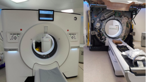
Urgent cases had same-day CT access, while PET imaging, though available, was less frequently used. For cardiac evaluation, a 256-detector CT scanner with 16 cm detector coverage and 0.23 mm slice thickness was employed.
In cases of suspected tricuspid endocarditis, a modified protocol was used to optimize imaging of the right heart chambers. Secondary acquisitions in the venous phase were routinely performed in complex cases to enhance visualization of perivalvular structures and associated complications.
Data were collected retrospectively from patients diagnosed with infective endocarditis between October 2024 and March 2025.
Technical Protocol
- Non-contrast Acquisitions: The initial non-contrast acquisitions were performed to evaluate coronary and extracoronary calcifications. These scans are essential for differentiating aneurysms and pseudoaneurysms from postsurgical changes and identifying complications such as intramural hematomas. Atypical calcifications, such as those observed in the mitral annulus, were also examined for their distinctive morphology and potential diagnostic challenges. A notable example is the caseous necrosis of the mitral valve "toothpaste tumor," a rare form of calcification that can mimic infectious or neoplastic processes, necessitating careful differentiation.
- Contrast Protocol: Contrast-enhanced imaging optimized visualization of perivalvular structures and associated complications, leveraging multiphasic protocols and high-resolution reconstructions for improved diagnostic accuracy. A contrast agent with a concentration >350 mg/ml was administered in volumes of 70-100 ml, adjusted for patient weight, with a flow rate of 5 ml/s, followed by a saline flush. In cases of suspected tricuspid endocarditis, a modified protocol was used to optimize imaging of the right heart chambers. Secondary acquisitions in the venous phase were routinely performed in complex cases to enhance visualization of perivalvular structures and associated complications.