Congress:
ECR25
Poster Number:
C-23763
Type:
Poster: EPOS Radiologist (scientific)
Authorblock:
A. Giri, P. C. P. Joshi; Pune/IN
Disclosures:
Ashana Giri:
Nothing to disclose
Priscilla Col Priscilla Joshi:
Nothing to disclose
Keywords:
Abdomen, Kidney, Urinary Tract / Bladder, CT, Diagnostic procedure, Calcifications / Calculi
This was a prospective observational study conducted on 70 patients over a period of 18 months.
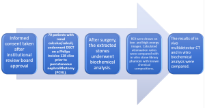
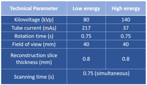
Fig 4: Approach to acquire DECT in our institute
DECT scans were performed using dual-energy protocols at 140 kVp and 80 kVp.
The attenuation profiles of various calculi (calcium oxalate, uric acid, cystine, and mixed composition) were analyzed and compared with biochemical analysis post-surgical extraction.
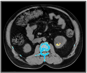
Fig 6: 60 yr/M, complained of left flank pain. No history of previous surgery or hematuria.
Overlay of high and low energy image.

Fig 7: Region of interest (ROI) drawn.
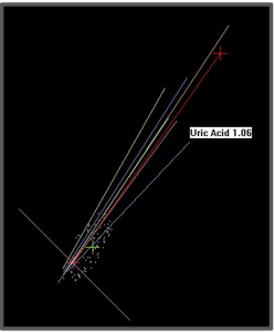
Fig 8: DECT spectral analysis showed uric acid, confirmed by biochemical analysis.