The white matter is organized into three distinct functional groups
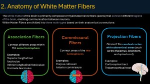
These tracts connect different cortical areas within the same hemisphere, enabling complex cognitive tasks.
-
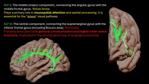
Superior Longitudinal Fasciculus (SLF): A massive bundle divided into three segments (SLF I, II, and III). It is the backbone of the "Where" visual pathway and visuospatial attention.
, ,
, .
.
-
Arcuate Fasciculus: Historically the most famous tract. It connects the Broca’s area (motor speech) with Wernicke’s area (understanding).
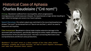
A lesion here leads to conduction aphasia. A poignant historical example is the French poet Charles Baudelaire, who, following a stroke that severed this "bridge," lost his ability to form sentences, his vocabulary tragically reduced to the repetitive exclamation: "Sacre Bleu!"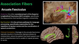
-
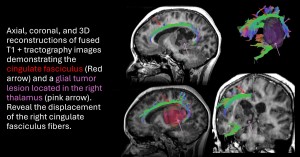
Cingulate Fasciculus (Cingulum): Curving over the corpus callosum, it is the primary tract of the limbic system, connecting the frontal lobe to the hippocampus, essential for memory and emotion.
, .
.
-
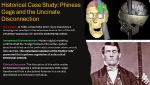
Uncinate Fasciculus: A hook-shaped tract connecting the orbitofrontal cortex to the anterior temporal lobe.
This tract gained fame through the case of Phineas Gage. When a tamping iron destroyed this connection, Gage’s executive "city" (frontal lobe) could no longer regulate his emotional "city" (limbic system), transforming his personality from a reliable foreman to an irreverent, impulsive man.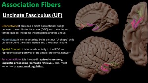
-
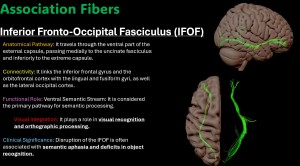
Inferior Fronto-Occipital Fasciculus (IFOF): The longest association tract, serving as the "What" stream for semantic processing.
-
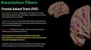
Frontal Aslant Tract (FAT): A more recently mapped tract connecting the Supplementary Motor Area (SMA) to Broca’s area, crucial for speech initiation.
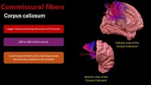
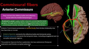
These fibers ensure that the two halves of the brain work as a single unit.
-
Corpus Callosum: With over 200 million axons, it is divided into the rostrum, genu, body, and splenium. Its fan-like radiations (Forceps Minor and Major) are easily identified on DTI by their vibrant red color (indicating left-right orientation).
-
Anterior Commissure: A smaller but vital bridge for olfactory and temporal lobe communication.
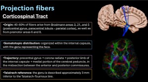
These tracts connect the cortex to subcortical structures, the brainstem, and the spinal cord.
-
Corticospinal Tract (CST): The most critical motor pathway. It descends from the motor cortex through the posterior limb of the internal capsule. In neurosurgery, mapping the CST is vital to prevent post-operative hemiparesis.
-
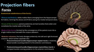
Fornix: The main output tract of the hippocampus, following a complex C-shaped trajectory around the thalamus.
-
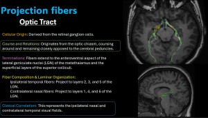
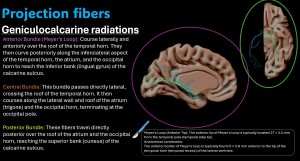
Optic Radiations (Geniculocalcarine Tract): Carries visual information to the occipital lobe. It includes Meyer’s Loop, which travels far forward into the temporal lobe. Radiologists must highlight this tract during temporal lobe epilepsy surgeries to avoid causing "pie in the sky" visual field deficits.