
The study of white matter has transitioned from gross dissection to sophisticated digital reconstruction. While Andreas Vesalius in the 16th century provided the first glimpses into the brain’s internal structure, it was Thomas Willis in the 17th century who revolutionized our understanding. Willis moved beyond the idea of the "ventricular soul" to propose that the white matter was a functional tissue capable of transmitting signals. His meticulous dissections laid the groundwork for what we now recognize as axonal connectivity.

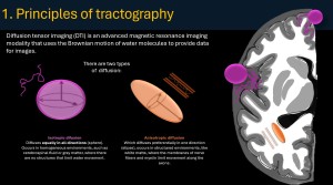
At the heart of modern white matter imaging is the Brownian motion
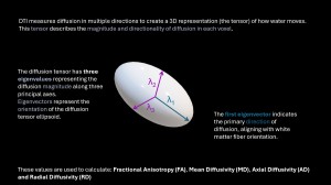
To capture this directionality, MRI sequences apply gradients in multiple directions (at least 6, though clinical protocols often use 30 or more). This data is used to calculate a 3D mathematical model called the Tensor, represented as an ellipsoid. From this tensor, we derive crucial quantitative metrics
-
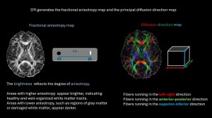
Fractional Anisotropy (FA): Measures the degree of directionality (0 for isotropic, 1 for perfectly linear). A drop in FA often indicates loss of structural integrity
-
Mean Diffusivity (MD): Reflects the overall presence of obstacles to diffusion
-
Axial (AD) and Radial Diffusivity (RD): Provide insights into axonal damage versus demyelination, respectively
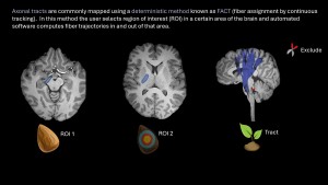
Tractography is the process of visually "weaving" these tensors into continuous lines. It begins with the placement of a Seed, a starting point or Region of Interest (ROI) from which the computer begins to track diffusion vectors.
-
Deterministic Tractography: Follows the primary eigenvector from voxel to voxel. If the angle between tensors is too sharp or FA is too low, the tracking stops. It is fast and clean but struggles with "crossing fibers"
-
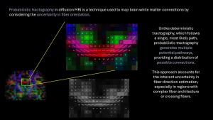
Probabilistic Tractography: Accounts for uncertainty in each voxel, creating a "probability map" of connections. This is superior for complex areas where different tracts intersect.
-
Multimodal Fusion: The integration of tractography with structural sequences like FLAIR or T1-weighted images allows the radiologist to see how a lesion (e.g., a tumor) interacts with the tract—whether it is displaced, infiltrated, or completely disrupted