Tumor Characteristics
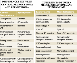
- Central Neurocytoma: Commonly located near the foramen of Monro, typically attached to the septum pellucidum, central neurocytomas display a characteristic "bubbly" appearance on T2-weighted MRI. Calcifications and cystic components contribute to their heterogeneous morphology. These tumors typically show intermediate T1 signal intensity and subtle post-contrast enhancement. [2,6]
 Fig 1: 25 year old male presented with history of intermittent headache for 1 month. MRI showed a well-defined lesion with irregular margins in the body of right lateral ventricle extending into foramen of Monro & 3rd ventricle which is isointense on T1W (Image A) & T2W (Image B), with diffusion restriction, SWI blooming (Image D), mild patchy contrast enhancement (Image C) and causing obstruction of foramen of Monro with hydrocephalus. MRS shows increased choline within the lesion. MR perfusion shows increased perfusion. -- Suggestive of Central Neurocytoma. Right frontal craniotomy and excision of mass was done. HPE was consistent with Central Neurocytoma, WHO grade II tumor. Follow-up CT after 6 months showed smaller recurrent/residual lesion, with calcifications within the lesion, but resolution of hydrocephalus (Image E). Image courtesy: Ananthapuri Hospitals and Research Institute, Thiruvananthapuram, Kerala, India.
 - Ependymoma : Observed predominantly in pediatric patients, ependymomas typically involve the posterior fossa and the fourth ventricle. On MRI, these tumors exhibit mixed solid-cystic features, hypointense T1 signals, and hyperintense T2 signals. CT often reveals calcifications and intratumoral hemorrhages. Post-contrast enhancement patterns are heterogeneous. Posterior fossa ependymomas often extend in a “plastic” way through the foramina of Luschka and/or Magendie. Hydrocephalus is a common complication requiring timely surgical intervention. [1,6]
 Fig 2: 32 year old male presented with altered behavior and irrelevant talk. MRI showed a heterogeneous lesion seen in the floor of IVth ventricle extending through the foramen of Magendie into the upper cervical canal and into the cisterna magna. The lesion is hypointense on T1W (Image A) & hyperintense on T2W (Image B and C) with blooming (Image E), heterogeneous enhancement (Image D) and no diffusion restriction. MR spectroscopy shows increased choline. MRI whole spine showed no evidence of lesions. -- Suggestive of Ependymoma. Posterior fossa surgery done and tumor excised. HPE was consistent with Ependymoma, WHO grade II tumor. Image courtesy: Ananthapuri Hospitals and Research Institute, Thiruvananthapuram, Kerala, India.
 - Medulloblastoma : Primarily affecting children, medulloblastomas are hypercellular tumors confined to the fourth ventricle. The growth is often rapid and accounts for their relatively rapid clinical onset. MRI frequently shows restricted diffusion on DWI and demonstrate heterogeneous contrast enhancement. These tumors often obstruct CSF flow, necessitating urgent ventriculoperitoneal shunting followed by resection. [4,6]
 Fig 3: 10 year old boy with vomiting and pain over neck. Well-defined lobulated lesion seen in IVth ventricle which is hypointense on T1W (Image A) & hyperintense on T2W (Image C) with diffusion restriction & patchy contrast enhancement (Image B). Multiple enhancing lesions seen in bilateral cerebellar hemispheres. Visualized spinal cord shows intramedullary lesion from the level of C1 to D4 with multiple cystic areas. Multiple enhancing dural based lesions seen posteriorly in the spinal canal (Image D). –Suggestive of medulloblastoma with spinal metastases. Suboccipital craniotomy with tumor excision done. HPE consistent with Medulloblastoma, WHO grade IV. Image courtesy: Ananthapuri Hospitals and Research Institute, Thiruvananthapuram, Kerala, India.
 - Glioblastoma Multiforme : Rare intraventricular glioblastomas exhibit aggressive features, including marked enhancement, necrotic centres, and peritumoral edema. These tumors are iso to hypointense to grey matter on T1, iso to hyperintense to grey matter on T2 and demonstrate variable heterogeneous contrast enhancement due to necrosis and microhemorrhages. MRS reveals increased choline, lipid and lactate peaks. Multimodal treatment with surgery, radiotherapy, and chemotherapy is required, though prognosis remains poor. [2,5]
 Fig 4: 42 year old male with headache. MRI shows heterogeneously enhancing (Image B), T1 hypointense and T2 hyperintense lesion (Image A) within the 3rd ventricle with infiltration of midbrain posteriorly. No diffusion restriction or blooming. MR Spectroscopy shows elevated choline (Image C). Tumor excision done and HPR came as grade IV glioma. Follow up MRI after 2 yrs shows recurrence in left posterior aspect of midbrain (Image D and E). Image courtesy: Ananthapuri Hospitals and Research Institute, Thiruvananthapuram, Kerala, India.
 - Subependymal Giant Cell Astrocytoma (SEGA) : Associated with tuberous sclerosis, SEGAs are slow-growing benign tumors near the foramen of Monro. They are generally larger than 1 cm, show calcifications, heterogeneous pattern on T1 and T2 and intense contrast enhancement. They can be either asymptomatic or symptomatic due to obstructive hydrocephalus. Surgical resection yields excellent outcomes in most cases. [6,3]
 Fig 5: 31 year old male with history of seizure. MRI shows multiple cortical and subcortical tubers in bilateral fronto-parietal lobes (Image A and C). Multiple small subependymal nodules, which on CT correlation shows calcifications (Image D and E). Heterogeneously enhancing lesion in the region of right foramen of Monroe causing infiltration of anterior septum pellucidum (Image B). -- Suggestive of Subependymal giant cell Astrocytoma. Image courtesy: Ananthapuri Hospitals and Research Institute, Thiruvananthapuram, Kerala, India.
 - Pilomyxoid Astrocytoma : This rare tumor in young children shows a well-circumscribed solid-cystic appearance on MRI, with homogenous post-contrast enhancement of the solid component. Approximately 20% show intratumoral haemorrhage. Surgical resection is the primary treatment, with adjuvant therapy for residual disease. [1,2]
 Fig 6: 32 year old female presenting with headache. MRI shows lobulated heterogeneously enhancing intraventricular lesion (Image B) within the lateral ventricles in the midline involving the septum pellucidum and infiltration of right basal ganglia with surrounding edema with associated hydrocephalus due to obstruction at the foramen of Monroe (Image A). Factors favoring intraventricular glioma includes involvement of basal ganglia and heterogeneous contrast enhancement. As the lesion is well defined and lobulated, possibility of low grade astrocytoma was also considered. HPR was consistent with pilomyxoid astrocytoma. Image courtesy: Ananthapuri Hospitals and Research Institute, Thiruvananthapuram, Kerala, India.
 - Metastases : Intraventricular metastases, though rare, are more common in older adults. Their imaging characteristics depend on the primary malignancy. MRI often shows heterogeneous lesions with variable enhancement. [5,4]
 Fig 7: 61 year old male. MRI showing heterointense lesion in the atrium of left lateral ventricle extending into the temporal horn (Image A). The lesion shows heterogeneous enhancement with subependymal enhancement extending into the occipital horn (Image B). There is adjacent brain parenchymal infiltration with extensive disproportionate edema. MR spectroscopy showed lipid lactate peak which is characteristically seen in metastases (Image C). CT guided biopsy was done for this patient which showed atypical cells, consistent with metastases from small cell neoplasm. Subsequent CT chest revealed evidence of bronchogenic carcinoma. Image courtesy: Ananthapuri Hospitals and Research Institute, Thiruvananthapuram, Kerala, India.
 - Choroid Plexus Papilloma and Carcinoma : Papillomas are benign, vascular tumors common in pediatric lateral ventricles, while carcinomas are malignant counterparts seen in adults. Both present with vivid contrast enhancement and can cause hydrocephalus due to CSF overproduction or obstruction. [3,5]
 Fig 8: 18 year old female. MRI showed a well-defined mildly lobulated intensely enhancing lesion in the atrium of left ventricle which is seen arising from the choroid plexus (Image D and E). The lesion is isointense on T1W (Image A) and hyperintense on T2W/FLAIR (Image B and C). There is mild dilatation of atrium of left ventricle. No evidence of hydrocephalus. There is no extension of the lesion outside the ventricle. No evidence of diffusion restriction/SWI blooming within the lesion. --Suggestive of Choroid plexus papilloma. Image courtesy: Ananthapuri Hospitals and Research Institute, Thiruvananthapuram, Kerala, India.
 Fig 9: 6 year old girl presented with history of seizure. MRI showed a large heterogeneously enhancing lobulated intraventricular lesion with the epicenter in the trigone of left lateral ventricle and causing significant midline shift (Image C). There is infiltration of brain parenchyma with the lesion reaching upto the dura with scalloping of the calvaria and transtentorial herniation. Mild hydrocephalus involving bilateral lateral ventricles. The lesion is predominantly solid which is isointense on T1W (Image A) & T2W (Image B) and highly vascular with the prominent draining vein which is seen draining into the left internal cerebral vein & vein of Galen. Multiple necrotic areas seen within. Mild diffusion restriction seen in the solid areas. MRS shows increased choline. MR perfusion shows increased perfusion. No leptomeningeal contrast enhancement or evidence of drop metastasis. --Suggestive of Choroid plexus carcinoma. Image courtesy: Ananthapuri Hospitals and Research Institute, Thiruvananthapuram, Kerala, India.
 - Colloid Cyst : These benign lesions are frequently found at the foramen of Monro and can intermittently obstruct CSF flow, causing episodic headaches. MRI reveals hyperintense lesions on T1-weighted images with rarely a thin rim enhancement, probably due to enhancement of the adjacent and stretched septal veins. Surgical excision is curative. [4,3]
 Fig 10: 44yr old male presented with headache. MRI showed a cyst hyperintense on T1W (Image A), heterointense on T2W (Image B) with blooming (Image C) located anterior to third ventricle in the region of foramen of Monroe. Margins appear irregular suggestive of contained rupture. There is mild hydrocephalus involving bilateral lateral ventricles with mild periventricular CSF seepage. Correlative CT confirmed the same (Image D and E). --Suggestive of colloid cyst. Right FP craniotomy was done and transcortical excision of hemorrhagic cyst was done. Image courtesy: Ananthapuri Hospitals and Research Institute, Thiruvananthapuram, Kerala, India.
Diagnostic Challenges
Diagnosing intraventricular tumors presents notable complexities due to overlapping imaging features, which can blur the distinctions among tumor types.
For instance, both central neurocytomas and subependymomas often appear as well-demarcated masses situated near the foramen of Monro. Differentiating between these entities requires careful analysis of imaging characteristics, such as signal intensities on MRI sequences and the presence of calcifications or cystic components. Similarly, dense enhancement patterns observed in tumors like meningiomas and choroid plexus carcinomas necessitate a comprehensive evaluation of the clinical context, including the patient's age and symptoms, as these factors provide critical diagnostic clues. For example, choroid plexus carcinomas are more common in pediatric patients, whereas meningiomas are typically seen in older adults. Advanced imaging modalities, including diffusion-weighted imaging (DWI), perfusion imaging, and spectroscopy, can offer additional insights but often require expert interpretation. These diagnostic challenges underscore the importance of integrating radiological findings with clinical data to establish a precise diagnosis and guide management strategies effectively.
Management Strategies
The management of intraventricular tumors was individualized, emphasizing a multidisciplinary approach tailored to the specific tumor type, patient demographics, and clinical presentation. Surgical resection emerged as the cornerstone of treatment, particularly for tumors causing significant mass effect or obstructive hydrocephalus, where prompt intervention was necessary to alleviate symptoms and prevent further complications. For benign tumors like subependymomas or central neurocytomas, gross total resection often sufficed, resulting in favorable outcomes. However, for malignant lesions such as glioblastomas or choroid plexus carcinomas, additional modalities like radiotherapy and chemotherapy were indispensable components of the treatment regimen. Radiotherapy was particularly valuable for controlling residual disease post-surgery, while chemotherapy played a role in addressing disseminated or recurrent malignancies. The collaboration between radiologists, neurosurgeons, and oncologists was pivotal in ensuring optimal care. This team-based approach facilitated accurate diagnosis, precise surgical planning, and the integration of adjunctive therapies, ultimately improving patient outcomes. Emerging technologies, such as intraoperative MRI and advanced neuronavigation systems, also enhanced surgical precision, further underscoring the dynamic and evolving nature of intraventricular tumor management.