Congress:
ECR24
Poster Number:
C-13414
Type:
EPOS Radiologist (educational)
DOI:
10.26044/ecr2024/C-13414
Authorblock:
A. Gavrilovici; Cluj Napoca/RO
Disclosures:
Anamaria Gavrilovici:
Nothing to disclose
Keywords:
Abdomen, Liver, CT, MR, Diagnostic procedure, Cirrhosis
The caudate lobe becomes enlarged through a compensatory mechanism, being a common finding in chronic liver diseases, and it is often associated with other morphologic abnormalities. While these changes are not pathognomonic, they can represent a piece in the diagnostic puzzle. Clear understanding of the pathophysiological process, corroborated with other key imaging findings, can help radiologists use the caudate lobe size in this differential diagnosis.
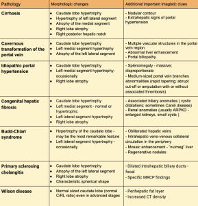
Table 5: Main pathologies associated with caudate lobe hypertrophy and abnormal liver morphology.
Additional imagistic features important for diagnostic.