Congress:
ECR24
Poster Number:
C-22236
Type:
EPOS Radiologist (educational)
DOI:
10.26044/ecr2024/C-22236
Authorblock:
A. A. Basheer Ahmed1, K. Saravanan1, P. G. Elangovan1, V. Adhithiya1, F. Abubacker Sulaiman2; 1Melmaruvathur, Tamil Nadu/IN, 2Chennai/IN
Disclosures:
Ashraf Ahmed Basheer Ahmed:
Nothing to disclose
K Saravanan:
Nothing to disclose
Prem Gowtham Elangovan:
Nothing to disclose
Vigneshwar Adhithiya:
Nothing to disclose
Farook Abubacker Sulaiman:
Nothing to disclose
Keywords:
Contrast agents, Catheter arteriography, CT-Angiography, PET, Barium enema, Outcomes
- Contrast media used with imaging techniques to enhance the differences seen between the body tissues on the images.
- Contrast media alter the response of the tissues to the applied electromagnetic or ultrasound energy by a variety of mechanisms.
- The ideal contrast medium would achieve a very high concentration in the tissues without producing any adverse effects. Unfortunately, so far this has not been possible, and all contrast media have adverse effects.
- Positive and negative contrast agents.
- The positive contrast media attenuate X-rays more than do the body soft tissues.
- Water soluble iodine agents
- Non-water-soluble barium agents.
- Negative contrast media attenuate X-rays less than do the body soft tissues. No negative contrast media are commercially available i.e air
- Barium sulphate preparations used to visualize the gastrointestinal tract consist of a suspension of insoluble barium sulphate particles which are not absorbed from the gut.
- Differences between the different commercially available agents are very minor and relate to the additives in the different barium sulphate preparations.
- Contain paramagnetic or superparamagnetic metal ions which affect the MR signal properties of the surrounding tissues.
- They are used to enhance contrast, to characterize lesions and to evaluate perfusion and flow-related abnormalities. They can also provide functional and morphological information.
- The most widely used paramagnetic contrast agents are non-specific extracellular gadolinium chelates.
- Their active constituent is gadolinium, a paramagnetic metal in the lanthanide series, which is characterized by a high magnetic moment and a relatively slow electronic relaxation time.
- Non-specific extracellular gadolinium chelates can be classified by their chemical structure, macrocyclic or linear, and by whether they are ionic or nonionic.
- also include liver specific gadolinium-based agents (gadobenate dimeglumine, Gd-BOPTA and gadoxetate, Gd-EOBDTPA) and manganese-based preparations [manganese chelate (mangafodipir trisodium) and free manganese combined with vitamins and amino acids (to promote the uptake) for oral intake]
- Superparamagnetic contrast agents include
- Superparamagnetic iron oxides (SPIOs).
- Ultra small superparamagnetic iron oxides (USPIOs).
- Two preparations of SPIOs are available: ferumoxides and ferucarbotran.
- These particulate agents are composed of an iron oxide core, 3–5 mm in diameter, covered by low molecular weight dextran for ferumoxides and by carbodextran for ferucarbotran.
- SPIOs are approved for liver imaging and
- USPIOs are under consideration for MR lymphography.
- After injection, SPIO and USPIO particles are metabolised into a soluble, non superparamagnetic form of iron. Iron is incorporated into the body pool of iron (e.g. ferritin, hemosiderin and hemoglobin) within a few days.
- Ultrasound contrast agents produce their effect by increased back-scattering of sound compared to that from blood, other fluids and most tissues.
- Ultrasound contrast agents can be divided into five different classes:
Radiographic Contrast Media:
Barium Contrast Media:
Iodinated contrast media:
Osmolarity –
High osmolar, low osmolar, iso-osmolar
Ionic state –
ionic and non-ionic
No. of benzene rings –
monomeric and dimeric
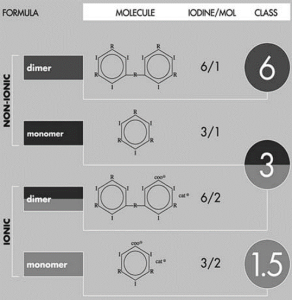
Fig 1: Iodinated Contrast Media
Magnetic resonance (MR) imaging contrast agents:
Ultrasound contrast media:
i.Nonencapsulated gas microbubbles (e.g. agitated or sonicated)
ii.Stabilized gas microbubbles (e.g. with sugar particles)
iii.Encapsulated gas microbubbles (e.g. by protein, liposomes or in polymers),
iv.Microparticle suspensions or emulsions [perfluorooctyl bromide (PFOB), phase-shift],